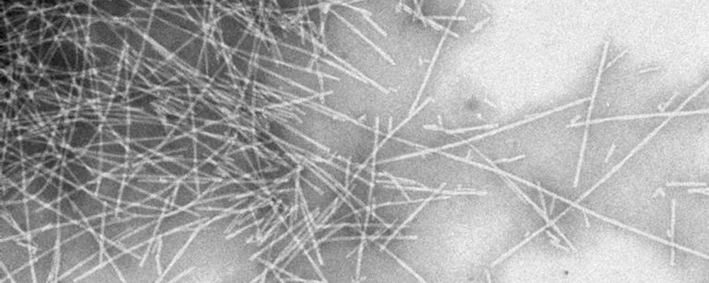
Negative staining
Negative staining is a rapid method for screening the morphology, size and arrangement of particulate samples such as proteins, vesicles, isolated organelles, whole viruses as well as bacterial flagellae and pili. The technique can be combined with immunogold detection.
The negative staining technique requires very little specialized equipment and sample suspension can be mounted directly on TEM support grids and examined after air-drying. However, due to their small size and low atomic numbers of the constituent atoms, the particles are relatively electron-lucent, i.e. they do not cause considerable electron scattering when they interact with the TEM beam. This means that they have very low contrast and do not stand out from the background.
In order to visualize particulate samples a heavy metal stain such as uranyl acetate or phosphotungstic acid is used. The particles are surrounded by the stain, which scatters electrons strongly creating negative contrast, whereby the particle appears bright against the stain. This is the opposite of the stain binding to the sample and excess being removed, in which case a particle appears dark against bright background. Parametres such as staining time and blotting technique affect the proportions between negative and positive staining on the grid and may have to be optimized for each sample type.

