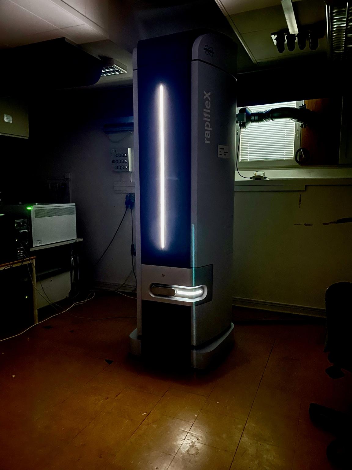
MALDI Imaging Mass Spectrometry
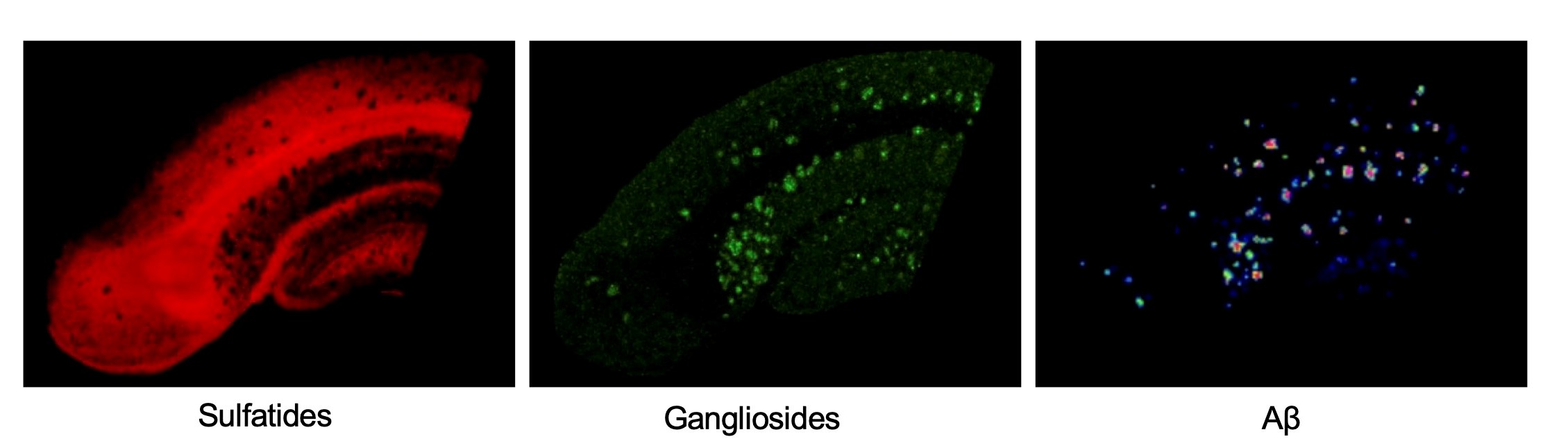
MALDI imaging mass spectrometry (IMS) is chemical imaging of lipids, endogenous peptides and small proteins with 5um spatial resolution. The image above shows localization of lipids in the brain. Left: Heatmap of sulfatide ST18:0. Right: Overlay image of sulfatide ST18:0 (red) and ganglioside GM1 (green).
For MALDI Imaging, CCI will provide advanced expertise for study design, feasibility and sample collection as well as for correlative imaging with other bioimaging techniques including immunohistochemistry and histology.
The staff at CCI will provide the whole IMS service from sample preparation to data acquisition and initial data analysis. Following an initial project meeting, the user can decide whether to start with a pilot project to further establish feasibility and hypothesis generation for a fixed price or proceed with a more large-scale study at costs/sample.
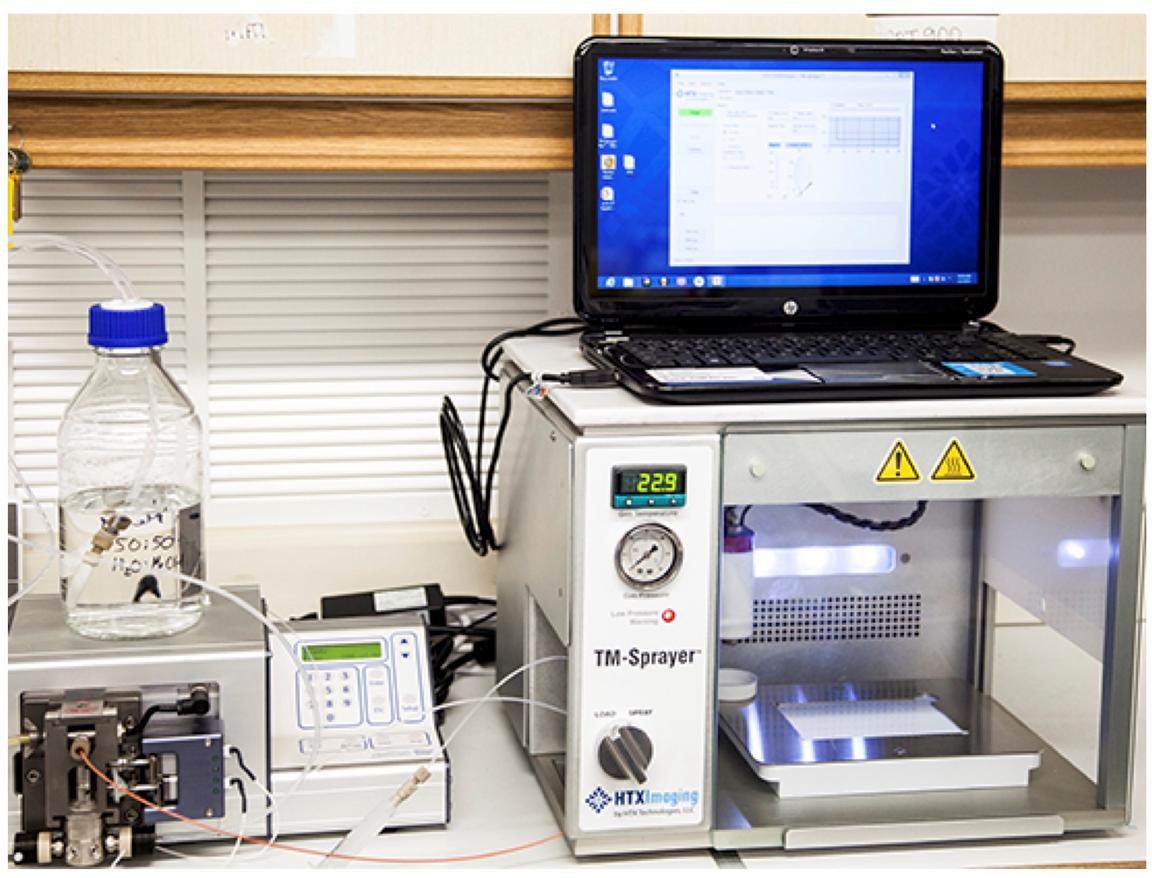
For each experiment, the user will provide fresh frozen specimen that can be processed for imaging of metabolites, lipids, endogenous peptides and small proteins.
In addition, to MALDI IMS, CCI will assist in planning and performing complementary microscopy experiments from the same or adjacent tissue sections.
Further, CCI will provide expertise for appropriate image analysis and statistical methods to investigate the generated imaging data.
NOTE: This full service is subject to feasibility as well as availability of our resources, and the pricing needs to be negotiated based on sample size and types of requested analyses. For more information please see MALDI-IMS prices and/or contact us.
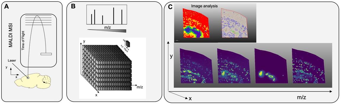
The principle of MALDI imaging MS (view larger image): (A) Tissue cryosections are mounted on a target for MSI. MALDI MSI involves coating of the tissues with matrix for desorption and ionization upon irradiation with a UV laser. MALDI facilitates softer ionization without extensive in source fragmentation and allows detection of large lipids, peptides and proteins. (B) MALDI MSI is performed by stepwise, systematic scanning with the probe and MS data acquisition based on a pre-defined array. The pixel-to-pixel distance defines the spatial resolution. (C) MALDI MSI data are interrogated comprehensively through multivariate image analysis. This reveals pathology specific chemical localization patters as exemplified for plaque pathology in Alzheimer’s disease. Single ion images are then generated for the distinct chemical species localizing to pathological regions and features. Ion images are generated by visualizing the intensity distribution of individual ion signals (m/z; Intensity) over the tissue array.
