Events at CCI
The CCI regularly organizes conferences, workshops and seminars in collaboration with other universities in Sweden and abroad and with industrial partners
CALL4HELP: Monthly event
CALL4HELP in Light Microscopy, Image Acquisition & Analysis
Are you struggling with your imaging pipeline and need advice on which microscopy technique is best for your sample, what analysis workflow would best answer your question or simply how to set up the whole imaging experiment?
CCI can help!
On the 3rd Friday of each month we are organising open hours (13:00 – 15:00) where our light microscopy and bioimage analysis experts can meet with you and discuss your sample preparation protocol (if you share it in advance), which microscope to use, which microscopy modality to use (e.g. functional microscopy), how to generate appropriate controls, how to perform quantitative image analysis, and other topics.
To register follow the link to the Registration Form
- 2023 - Jan - 20th: In this meeting, participants brought questions related to image analysis and quantification. One was related to morphological descriptors (e.g., shape characteristics), and the other was related to intensity (level of expression) and spatial distribution.
Registration:
Where do we meet?
We meet in the computer room located at the light microscopy corridor of the CCI. Medicinaregatan floor 1, next to the elevator close to entrance 1F
The Centre for Cellular Imaging (CCI) is pleased to announce the purchase and installation of two new systems: A Super-resolution and a Laser Scanning Microscope for fast live imaging!!
Event Timing: | February 23rd, 2023 |
Lectures Address: | Hörsal Arvid Carlsson, Academicum, Medicinaregatan 3 |
Demo Address: | Medicinaregatan, floor 1, staircase of door 7A |
ZEISS ELYRA 7 with Lattice SIM² and Single Molecule Localization - Your Live Imaging System with Unprecedented Resolution. The super-resolution microscope ELYRA 7 with Lattice SIM² you can now double the conventional SIM resolution and discriminate the finest sub-organelle structures, even those no more than 60 nm apart. You don‘t need to sacrifice resolution when imaging at high speed (up to 255 fps ) using only the minimal exposure needed for life observation. ELYRA 7 enables you to combine super-resolution and high-dynamic imaging – without the need for special sample preparation or expert knowledge of complex microscopy techniques.
ZEISS LSM 980 with Airyscan 2 - Your Unique Confocal Experience for Fast and Gentle Multiplex Imaging. To analyze life with as little disturbance as possible, you must use low labeling density for your biological models. This requires excellent imaging performance combined with low phototoxicity and high speed. LSM 980, your platform for confocal 4D imaging, is optimized for simultaneous spectral detection of multiple weak labels with the highest light efficiency.
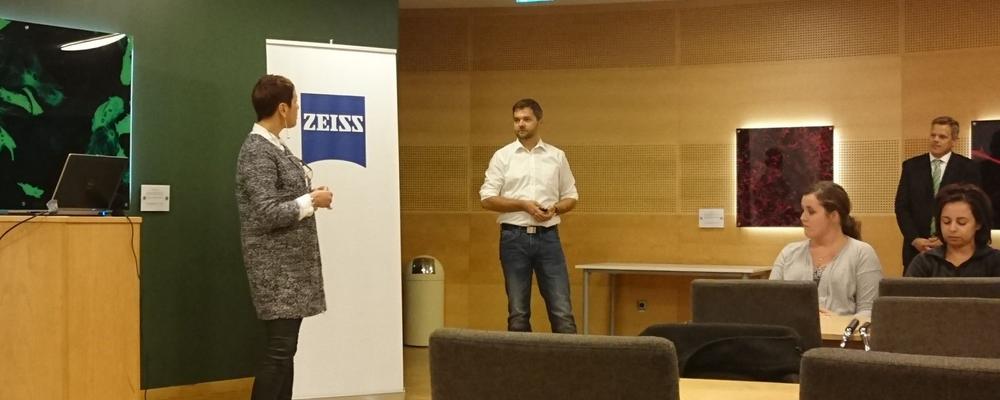
These two microscopes are now installed at the Centre and to celebrate this, we will host an “Opening day” with seminars and demos where you can learn about these advanced technologies that powers these class-leading instruments. During this day we would like to invite you to take a glimpse of what is possible with these microscopes, and what type of questions can be addressed.
Please, join us at the ”CCI Opening day”, 23rd of February 2023, to explore this new microscopes!
Register to by February 20th: link for registration
Venue for the lectures: Hörsal Arvid Carlsson, Academicum, Medicinaregatan 3 Google Maps link
Schedule
Time | Event |
---|---|
9:50 - 10:00 | Welcome from the Head of the Centre for Cellular Imaging, Dr Julia Fernandez-Rodriguez |
10:00 - 10:35 | LSM 980 with Airyscan 2 – Your Unique Confocal Experience for Fast and Gentle Multiplex Imaging. Dr Chris Power, Product & Application Sales Specialist, ZEISS Research Microscopy Solutions |
10:35 - 11:10 | ELYRA 7 – Ways to improve decoding algorithms in Structured Illumination and Localization Microscopy. Dr Klaus Weisshart, Product Management, ZEISS Research Microscopy Solution |
11:10 - 11:40 | The Arivis Scientific Imaging Platform: Analysis without Compromises, Molly McQuilken, Business Development Manager, Arivis at ZEISS company |
12:00 - 13:00 | Lunch Break |
13:00 - 15:00 | Visit to the CCI Facility and demos |
Open lectures, 18th of May - 21th of May, 2021: Artificial intelligence and bio-image analysis
As part of the "Smart Microscopy" workshop, see above, we have invited external speakers to give lectures on relevant topics in artificial intelligence and bio-image analysis (Deep Learning, Tissue analysis, Quantitative microscopy, Big Data handling, etc.). On this occasion, we are very fortunate to welcome:Carolina Wählby (SciLifeLab - Uppsala University, Sweden), David Dang (London's Global University - UK) and Jean-Yves Tinevez (Institut Pasteur, France). These lectures are open to anybody interested in these topics but registration is mandatory.
- Monday 17th of May, 13:00-13:30 CEST: Aliaksandr Halavatyi, "Designing adaptive feedback microscopy pipelines and performing experiments with AutoMicTools library in Fiji"
Lecture: https://youtu.be/7w9khqssZrw - Tuesday 18th of May, 13:00-13:30 CEST: David Dang, "SpinX: Time-resolved 3D Analysis of Spindle Dynamics using Deep Learning Techniques and Mathematical Modeling"
Lecture: https://youtu.be/7_SwQ4Cd1ME - Thursday 20th of May, 13:00-13:30 CEST: Carolina Wählby, "Image analysis and AI in microscopy-based life science research"
Lecture: https://youtu.be/EdiX1tJUSxk - Friday 21st of May, 13:00-13:30 CEST: Jean-Yves Tinevez, "Tracking cells for automated microscopy"
Lecture: https://youtu.be/mLUeSkpI07w
ONLINE opening day: Celldiscoverer 7 / LSM 900 Airyscan 2, 30 March, 2021
The Centre for Cellular Imaging is pleased to announce the purchase of an automated High Content Screening system combined with a Laser Scanning Confocal Microscope: the Celldiscoverer 7/ LSM 900 Airyscan 2 from Zeiss. With this system we open up the possibility for advanced automated live cell imaging with feedback experiments and guided acquisition including advanced image processing and analyzing capability! For more information, visit the Zeiss web page
This microscope is now installed at the CCI and to celebrate this, we will host an “ONLINE opening day” with seminars where you can learn about the advanced technology that powers this class-leading instrument. During this day we would like to invite you to take a glimpse of what is possible with today’s advanced automated live cell imaging system, and what type of questions can be addressed.
Registration:
Register to juliafer@cci.sahlgrenska.gu.se by March 26th. On March 29th you will receive the link for the Seminar
Program of the day:
13:00 - 13:15 Welcome and introduction; Dr Julia Fernandez Rodriguez, Head of the Centre for Cellular Imaging, and Dr Jens Wigenius, Zeiss Regional Sales Manager
13:15 - 13:45 "Advanced automated Live cell imaging with Zeiss Celldiscoverer 7/LSM 900 Airyscan2”; Dr. Sören Prag, Zeiss Application specialist in automated and advanced imaging systems
13:45 - 14:00 Short-break
14:00 - 14:20 Life Sciences Applications; Dr. Sören Prag, Zeiss Application specialist in automated and advanced imaging systems
14:20 - 15:00 Q&A
15:00 - 16:00 Live virtual demonstration Zeiss Celldiscoverer 7/LSM 900 Airyscan2; Dr Rafael Camacho, Scientific Officer Centre for Cellular Imaging, and Dr Maria Trulsson, Zeiss Application specialist


2019
Open-day and Workshop: Zeiss Gemini450 Scanning Electron Microscope, 25-26 November, 2019
In collaboration with Zeiss, the Centre for Cellular Imaging (CCI) is organizing an Open-day and Workshop to introduce our new Zeiss Gemini450 Scanning Electron Microscope. In particular, we will introduce and discuss the 3D Array Tomography - Image Serial Sections Fast and Efficiently – with Nano-resolution on this new microscope!
During the Open-day there will be technical and scientific presentations and short demos during the afternoon of the 25th of November. And for those interested, we will run on the 26th of November a full workshop in 3D Array-Tomography - from Sample Preparation to Image Processing!
Workshop: Live Cell imaging in Super Resolution Mode, 11 November, 2019
The workshop “Live Cell imaging in Super Resolution Mode” focuses on the new ZEISS ELYRA 7.
Join us for an insight into the applications of instruments and software for live cell imaging and take advantage of this unique opportunity to learn how your work can benefit from a flexible platform for fast and gentle superresolution imaging. There will be hands on with comparison of old and new systems.
Conference: SCANDEM 2019, june 11-14
Welcome to SCANDEM 2019 - from Atoms to Complex Systems - the
70th Annual Meeting of the Nordic Microscopic Society!
Topics: Imaging across scales/multimodal Microscopy (Correlative microscopy, cryo-EM, X-rays, Imaging Mass Spectrometry, etc.); Vibrational & Nonlinear Microscopy; Live & Fast super-resolution microscopy – Frontiers in imaging of ultrafast processes; Molecular and Functional Imaging; Quantitative high spatial resolution spectroscopy; In situ Microscopy; Artificial Intelligence in big data analysis, and computational microscopy.
Workshop: Live Cell Imaging in Different Technologies, 13 March, 2019
The workshop “Live Cell Imaging in Different Technologies” focuses on ZEISS Celldiscoverer 7 and recently launched ZEISS ELYRA 7.
Program highlights of the day:
• Live cell imaging - From high content to high resolution
• Analyzing your live cell data - Benefit from Intellesis software and image analysis
• Visualization of your live cell data - Deconvolution of data and Arivis tec hnologies
• Q&A and case studies
2018
Workshop: Nanoimager from ONI, Nov 22-23, 2018.
The Nanoimages from ONI is a new generation of super-resolution microscope, which reveals biology in a new light. We can now image cells at the highest resolution, understand molecular interactions and dynamics and study the fundamentals of biology through single-molecule fluorescence.
The system will be presentated on Thursday Nov 22 at 9 am in Jörgen Lehman, Academicum. Then, it will be possible to get a short demo of the system or to try it with your own samples during a slightly longer session during Nov 22-23.
2017
Workshop: Tools and Techniques for 3D Light and Electron Microscopy: Dec 12, 2017 - Gothenburg
This is a collaboration between the Centre for Cellular Imaging at the University of Gothenburg and Carl Zeiss. Scanning electron microscopes (SEMs) are increasingly used in biomedical research to obtain large volume data of biological samples. With a SEM you are not limited to grid-mounted samples and it is possible to produce hundreds of serial sections and investigate automatically in the SEM. Furthermore, blocks of cells or tissue can be processed directly in the microscope to produce large 3D volumes of pre-selected target areas. Using the latest software, this functionality offers a straightforward integration with other microscopy techniques such as fluorescence, laser scanning, and X-Ray microscopy. Applying these modalities enables you to combine functional and ultra-structural information across length-scales.
Program highlights
- Introduction into SEM portfolio
- Basics of SEM imaging in Life Sciences
- 3D volume in modern SEM methods
- Multi SEM
- Orion and or XRM
- Correlative Microscopy Scientific customer presentation related to SEM
Workshop: “Opening day” at the Centre for Cellular Imaging arranged together with Thermo Fisher Scientific, 2017
The Centre for Cellular Imaging is pleased to announce the installation of a new transmission electron microscope (TEM): Thermo Scientific™ Talos™ L120C TEM. This microscope is now installed at the CCI and to celebrate this we will host an “Open-Day" with seminars where you can learn about the advanced technology that powers this class-leading instrument. During this event we would like to invite you to take a glimpse into what is possible with today’s transmission electron microscopy, both in theory and practice, and learn what type of questions it can address!
Program:
13:00-13:10 – Welcome from the Head of the CCI, Julia Fernandez-Rodriguez
13:10-13:45 – Latest Technology and Solutions Focusing on Your Needs: Thermo Scientific Talos L120C TEM, Guarav Sharma
13:45-14:30 – Let’s Prove It! From Microscope to Your Success! Joanne Lo
14:30-16:30 – Visit to the CCI facility, microscope demonstrations* and refreshments
Workshop: Optical microscopy in life science - from basics to state of the art, 2017
4th Workshop CYCLON-HIT on the 3rd-4th of May 2017 at the Wallenberg conference centre and Centre for Cellular Imaging. Local organizers: Marica Ericson (Centre for skin research at University of Gothenburg & Chalmers) and Julia Fernandez-Rodriguez (CCI, University of Gothenburg)
Workshop: Light-sheet imaging of large cleared specimen, 2017
The CCI together with Zeiss will organize a seminar followed up with a workshop about the Lightsheet Z.1 system. Demo sessions will take place on the 5th and 6th of April. Please, register for the workshop-seminar to Julia Fernandez-Rodriguez, at the CCI juliafer@cci.sahlgrenska.gu.se by March 20th, and for the demo please book for individual demonstrations in advance via jens.wigenius@zeiss.com
2016
Workshop: “Opening day” at the Centre for Cellular Imaging arranged together with Carl Zeiss Nordics, 2016
The CCI is pleased to announce the installation of the LSM 880-Airyscan microscope with the fast acquisition module! This is the first system installed in the Nordics with the Airyscan-fast acquisition capability!
Please join us at the CCI Open day, to explore this new microscope!
Lecture-Hall: Birgit Thilander (Academicum), Medicinaregatan 3A
Program:
13:30-13:45 Welcome from the Head of the Core Facilities, Professor Göran Larson, and the Manager of the CCI, Dr Julia Fernandez-Rodriguez
13:45-15:00 Seminar: Airyscan goes Fast: An innovative use of the new confocal detector enables low-noise imaging at high frame rates. Dr Uroš Kržič, Senior Application Consultant at Carl Zeiss Microscopy
15:00- Visit to the CCI Facility, Refreshments!
Bridging Nordic Imaging Symposium, 2016
The CCI organized the Bridging Nordic Imaging Symposium and the 1st Cellular Imaging workshop, 14-15th of April 2016. Please, visit the BNI2016 homepage
2015
Demo system: LSM 800 Airyscan, 2015
ZEISS offers the possibility to test the LSM 800 Airyscan microscope at the Centre for Cellular Imaging. A demo system will be available in the facility during one week: 31st of August - 4th of September, 2015. Individual demo sessions can be booked via jens.wigenius@zeiss.com
Seminar: "LSM 8 family with SR Airyscan", 2015
ZEISS has recently launched the new LSM 880 and 800 with Airyscan. This opens a complete new world of confocal performance with fast and sensitive super resolution microscopy.
Seminar: March 30, 2015, at 13.30 in lecture hall "Carl Kylberg", Medicinaregatan 7B
Speaker: Hans Thorn, Application specialist, Carl Zeiss
Coffee will be served.
Please, sign up by e-mail to jens.wigenius@zeiss.com
Download full invitation
Workshop: STED superresolution microscopy, 2015
The Super-resolution stimulated emission depletion (STED) microscopy won the Nobel price in Chemistry 2014. Centre for Cellular Imaging now invites you to a seminar about this microscope and the underlying technique, STED! In addition, for those of you who would like to get a first look on your cells/tissue using this Super-resolution technique there is the possibility to sign up for a demonstration.
Seminar:
Tuesday March, 24, 2015, in lecture hall "Lyktan, Wallenberg Conference Center
9:00 Coffee
9:15 - 10:00 Microdedic & Leica Microsystems:
An introduction to Leica STED Super resolution Microscopy
10:00 - 11:00 Dr. Christoffer Lagerholm, Imaging facility manager, University of Oxford:
Applications of STED microscopy - Combining STED with singel molecule detection and particle tracking
Demonstrations:
Throughout the week the Leica TCS SP8 STED 3X system is installed at the Centre for Cellular Imaging.
For demos contact: Simen Gylterud Owe, Micromedic AB, simen.owe@micromedic.se, 47 91862041
For the workshop contact: Julia Fernandez-Rodriguez, Manager of the CCI, juliafer@cci.sahlgrenska.gu.se
Seminar and demonstrations: Quantitative Label-Free Live Cell Imaging, 2015
The Centre for Cellular Imaging invites you to a seminar and
demonstration of the Phasefocus VL21 label-free live cell imaging system.
Seminar:
Monday 23rd Febuary 2015 at 13:00 pm
Lecture Hall “Nils Wedel”, Medicinaregatan 9B.
Speaker: Joanne Whetstone, Phasefocus
Demonstrations:
Tuesday 24th - Friday 27th Febuary 2015. Please, sign up as soon as possible since there is a limited number of demo sessions.
Download full invitation
Please contact Maria Smedh, Centre for Cellular Imaging (maria.smedh@gu.se) or Joanne Whetstone at Phasefocus (joanne.whetstone@phasefocus.com) to book a demonstration.
Workshop: Atomic Force Microscopy, 2015
The Centre for Cellular Imaging Core Facility at the University of Gothenburg together with Bruker would like to invite you to a workshop in "Atomic Force Microscopy" (AFM). Discover the AFM technique, how it can be used for Biological applications and take the first step into the Nano World!
Date: Tuesday 9th December.
Lecture-hall: Ingegerd Eriksson, Medicinaregatan 3.
The aim of the workshop is to make you
10:00 - 12:00
Presentation made by Alexander Dulebo, Bruker Application engineer, in tight relation with biological Universities in Europe using AFM
1:00 – 17:00
One to one discussion with Bruker Biologist Expert
Register and book a time by contacting fabrice.magne@bruker-nano.com
2014
Seminar: Imaris, 3D image analysis software, 2014
Bitplane AG invites you to discover how the 3D image analysis software Imaris can help you analyse your data.
Speaker: Erik von Stedingk, Bitplane AG
Date: November 25, 2014
Time: 9:30-12:00
Location: Meeting room Curt Weibull (Medicinaregatan 3)
Seminar: "First Lab-on-a-tip technology in the world". This half-day seminar presents a multifunctional pipette for performing experimental cell biology at the single cell level.
Speaker: Gavin Jeffries, Asst. Professor at Chalmers University of Technology
Date: Thursday 16 October, 2014
Time: 9:45-12:00
Location: Meeting room Ingegerd Eriksson
(Academicum, the Sahlgrenska Academy, Medicinaregatan 3)
Download full invitation
Please sign-up your participation before Sept 30th by contacting:
Joanna Pylvänäinen, Centre for Cellular Imaging, or Gavin Jeffries, Chalmers University of Technology
Light-sheet microscopy seminar with Jacques Paysan, Zeiss Light-sheet application specialist, Germany.
24 of June at 14:00 in lecture hall Waldemar Sjölander
Open symposium "Super-resolution structured illumination and single molecule localization microscopy"
3rd of April 2014 in lecture hall "Lyktan" at Wallenberg Conference Center, Medicinaregatan 20A.
Organizers: Centre for Cellular Imaging and Zeiss. Detailed program
Workshop "Microfluidic platforms for functional cell studies - Live cell imaging to scan reality". 3rd of March 2014. Ingegerd Eriksson conference room, Academicum. Organizers: Centre for Cellular Imaging and Helena Wensman, Gradientech AB. For more information about the CellDirector visit the Gradientech webpage.
2013
Workshop "Using Microfluidics for Real-Time Imaging of In-Vitro Cell Models". September 2013. Organizers: Centre for Cellular Imaging and Merck Millipore. Click here for videos and product information.
2011
Workshop in a NEW Optical Sectioning Microscope. Revolution DSD, is an innovative white light confocal spinning disk based on ‘aperture correlation’. March 2011. Organizers: Centre for Cellular Imaging and Andor.
Volocity 3D Image Analysis Software Demonstration and Workshop. December 1, 2011, Birgit Thilander, Academicum, University of Gothenburg. Organizers: Centre for cellular Imaging and PerkinElmer
2010
Workshop on Non-contact Laser Capture Microdissection, February 2010. Organizers: Centre for Cellular Imaging and Carl Zeiss.
Pure sample preparation is an important prerequisite for specific and reliable molecular analysis. In our workshop we will show you an entirely non-contact method of cell or tissue isolation. Useful information regarding up- and downstream work will be addressed as well as demonstration of the laser microdissection microscope.
2008
Seminar on High‐Content Assay Development and Screening with Automated Imaging Systems, Image Analysis Tools, and Cellular Informatics Software. Seminar by Christian Holz Molecular Devices, June 2008. Organizers: Centre for Cellular Imaging and Molecular Devices.
Workshop on High Content Screening Microscopy, Scan^R Screening System, November 2008. Organizers: Centre for Cellular Imaging and Olympus.
2007
Miniseminar on Quantum Dots, presented by Stephen H. Chamberlain, March 2007. Organizers: Centre for Cellular Imaging and Invitrogen.
2006
Workshop on live cell imaging using conventional fluorescent microscopy, University of Gothenburg, 2006. Organizers: Centre for Cellular Imaging and Olympus.
Workshop on Laser Microdissection, Nov 2006. Organizers: Centre for Cellular Imaging and Leica Microsystems.
2005
Workshop on fluorescent microscopy, University of Gothenburg, March 2005. Organizers: Centre for Cellular Imaging and Leica Microsystems.
Workshop on fluorescent microscopy and Image analysis, University of Gothenburg June 2005. Organizers: Centre for Cellular Imaging and Olympus/Improvision.
2004
4th International Meeting of European Light Microscopy Initiative (ELMI). May 26-28, 2004 Gothenburg, Sweden.
Organizers: Tommy Nilsson, Maria Smedh and Julia Fernandez-Rodriguez, University of Gothenburg, Sweden.
Rainer Pepperkok, EMBL, Germany.
Anna Aragay and Jaakko Saraste, University of Bergen, Norway.
Workshop on fluorescent microscopy, University of Gothenburg, November 2004. Organizers: Centre for Cellular Imaging and Carl Zeiss.