Image gallery
Here are some examples of work performed by our users and the staff at CCI.

Top: Microvilli in mouse kidney imaged using Talos L120C. Courtesy of Roberto Boi.
Middle: TEM image of what is probably several nuclear pore complexes imaged by Charlotte Hamngren Blomqvist. With a little bit of imagination, it looks almost like an octopus swimming around inside the cell.
Bottom: A Golgi apparatus cluster in a HeLa cell prepared for serial block face-SEM, imaged using Talos L120C. Preparation and image: Anna Pielach


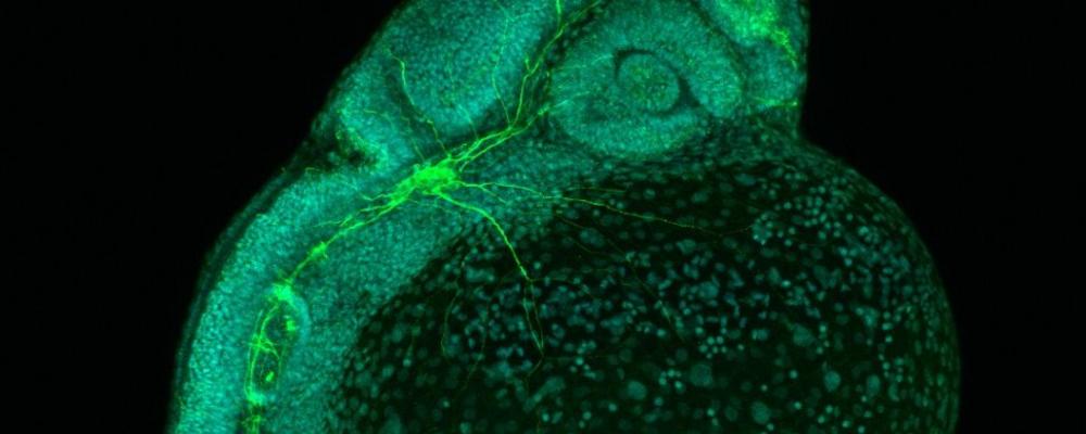
Top: Image of a zebra fish, acquired at the LSM700inv confocal microscope by Jasmine Chebli.
Middle: Zebrafish embryo imaged by Rakesh Banote using the LSM700inv confocal.
Bottom: A Barnacle larva. Sample by Anna Abramova imaged by Carolina Tängemo using DIC on the AxioObserver microscope.


Top: Superresolution image (i.e. SIM technology) by Carolina Tängemo, CCI, showing mitochondria stained with Mitotracker Red. Acquired at the Elyra PS.1.
Top (2): 3D multiphoton image of the penetration of sulforhodamine in normal skin. By Carl Simonsson, Department of Chemistry, Göteborg University.
Middle: HELA cells infected with Adenovirus 5 expressing green flourescent protein and the subunit B from Haemophilus ducreyi cytolethal distending toxin (HdCDT). The infected cells (green) are stained with DAPI (nuclei in blue) and an antibody to HdCDT (red). The image was acquired with the LSM 510 META microscope, using 63x/1.4 oil objective. By Teresa Lagergård and Julia Fernández-Rodríguez, Department of Microbiology, Göteborg University and CCI
Bottom: Collagen fibers in dermis layer from a human skin sample, seen by autofluorescence. The image was taken by Maria Smedh, CCI, using LSM710NLO.


During the 2nd Bridging Nordic Imaging Symposium in Gothenburg, CCI as local organizers, arranged the image competion "Art meets Science" where the conference participant were invited to create their own art work. The original image had to be a scientific image of a (biological) speciment, but otherwise any image processing technique or montage was allowed to give the image an extra dimension. At the exibition all participants could vote for their favorite based on how visually compelling and original they were.
Winner was the image "Cell Flair" submitted by Elnaz Fazeli, closely followed by "Cancer Scream" by Carolina Wählby and Leon A. Gatys (2nd place) and "Blossom" by Laure Plantard (3rd place).


Top: Salmon intestine section stained for sodium potassium ATPases. Red fluorophore shows an antibody staining several isoforms of the ATPase and green fluorophore an antibody specific against sodium potassium ATPase 1c. The tile image (2x4) was acquired on the AxioObserver widefield microscope using a 10x/0.3 objective. By: Jenny Lindström, Biological and Environmental Sciences.
Middle: Image displays the hind brain of 96 hrs old zebrafish embryos showing reticulospinal neurons (red) and motor neurons (green). Retrograde labelling was performed to trace neural connections in transgenic (gata2:GFP) 72hrs embryos, using rhodamine dextran, that was injected into the spinal cord. By: Rakesh Kumar Banote, Department of Psychiatry and Neurochemistry. Winner of the image competition.
Bottom: Dendrite from pyramidal neuron filled with Alexa-488. The picture was taken using super-resolution 3D-SIM microscopy on the ELYRA system, using 100x/1.45 oil objective, and reconstructed by surface 3D rendering with Zen2011 processing software. By: Marta Perez, Department of Neurophysiology.