
Grants for five Institute researchers from Lundberg Research Foundation
This year’s grants from the IngaBritt and Arne Lundberg Research Foundation were announced recently. Five of the recipients are researchers in clinical sciences, and they are jointly receiving more than SEK 21 million of the year’s total funding of SEK 37 million. As always, the grants go to research in cancer, kidney disease, and orthopedics.
This year's grants were awarded to research projects focusing on such areas as leukemia, kidney failure, metastatic cancer, and bone fractures. The Foundation prioritizes applications for the purchase of technical equipment for research. Geographically, the funds go mostly to projects in Gothenburg, Lund, and Stockholm. (Source: Lundberg Foundation.)
“Consistent themes are treatment with targeted therapies, AI in which instruments can recognize structures, and equipment enabling study of kidney transplantation outcomes. The Foundation's support is especially important since it’s often hard for researchers to obtain resources for investments in technical equipment,” says Olle Larkö, Professor of Dermatology and Venereology, and board member of the IngaBritt and Arne Lundberg Research Foundation.
"Treatment of metastatic cancers with radioactive drugs — development and testing of new strategies
Eva Forssell-Aronsson, Hospital Physicist and Professor of Radiophysics, is the recipient of the largest grant: SEK 13 million altogether. Her research is focused on side effects and cancer responses to radionuclide therapy.
“We’ve been awarded this funding to study new methods of treating metastatic cancer with radioactive tumor-seeking drugs. Primarily, we’re studying tumor diseases where the tumors express hormone receptors. The work comprises studies to boost the treatment effect on tumor tissue, and also methods to reduce side effects,” Forssell-Aronsson says.
The advantages of radioactive drugs are that they can reach dispersed metastases and tumor cells without their exact location in the body needing to be known, and that the radiation dose is delivered locally in the tumor tissue.
“The funds are for purchasing a preclinical PET/MRI system at the University of Gothenburg, for mouse or rat imaging. PET/MR equipment means that a magnetic camera and positron camera are combined, enabling an animal to be examined with both methods without needing to be moved.”
Abundant information in single system
MRI provides anatomical images, while PET produces functional ones. The combination of MR and PET shows where in the organ and/or tissue the parameters shown by PET are located. This allows the operator then to superimpose images from one camera on those from the other to obtain even more information. In addition, MRI can also give the scientists more functional information than PET provides.
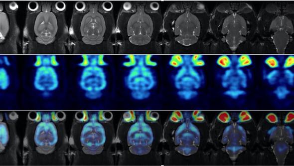
“This support for translational research in Gothenburg is a tremendous boon. We already had an outdated magnetic camera that’s been used in our project and many others in Gothenburg. The new PET/MR camera will be easier to use and provide images with superior performance. What’s more, it’s the first positron camera in Gothenburg for depicting animals. For patients, the first corresponding PET/MRI system has been installed at Sahlgrenska University Hospital, so now it’ll also be easier to study the same parameters both in animals and in patients.”
Other research groups too
The scientists will also engage in further work to develop methods of characterizing tissue better. The equipment will, moreover, be available to other research groups. This is the first time Forssell-Aronsson has applied for and received funding from the Lundberg Foundation.
Mechanical evolution of bone tissue around orthopedic implants
Anders Palmquist, researcher in the subject area of biomaterials, is receiving a grant of SEK 3 million for his research on bone-anchored (osseointegrated) amputation prostheses.
A femoral amputation impairs mobility and adversely affects both quality of life and well-being. Compared with a traditional sleeve prosthesis, an osseointegrated amputation prosthesis enhances both mobility and quality of life. Palmquist’s research has long addressed this treatment, based on an implant being allowed to heal into the bone and thus become firmly anchored there, and he has now received a grant of SEK 3 million from the Lundberg Research Foundation.
A conventional prosthesis, attached to the outside of the amputation stump by means of a prosthesis sleeve, restricts mobility, usually causes discomfort and gives rise to abrasions and sweating. A directly bone-anchored prosthesis can, instead, reduce these problems related to sleeve prostheses and make using a prosthesis easier.
“Much of the improvement is due to the load going straight to the bones instead of via the soft tissues. However, it requires the skeleton is adaptable to the ‘new’ forces generated by use of the prosthesis. In my research, I’ve developed several methods of studying how bone tissue grows, matures, and adapts around implants, and how this relates to mechanical function. This grant broadens our analytical platform, which is key for developing new, improved implants,” Palmquist says.
New atomic force microscope enabling study of nanomechanical properties
"Bone tissue is a nanocomposite material that adapts continuously to its mechanical and biochemical environment, with remodeling and maturation progressively enhancing mechanical proficiency.With the new atomic force microscope, we can study the nanomechanical properties of the bone tissue around implants, and correlate these properties with itslocal structure and composition,” says Furqan Ali Shah, a co-applicant and associate professor in the research group.”
“Experimental studies and continued follow-up of clinical implants after selectionhelp us understand how the mechanical evolution of bone tissue takes place, and how it’s affected by implant design, skeletal quality, load, and disease. This knowledge is used to optimize the implants of the future for treating patients with reduced skeletal quality and healing capacity,” Palmquist says.
His research group's focus is on seeing and clarifying the connection between biomechanical function on the one hand and, on the other, structure and chemical composition at the interface between implant surface and bone tissue.
Equipment for studying infections connected with orthopedic implants
Margarita Trobos, another associate professor in the field of biomaterials, is to receive SEK 1.7 million to fund "Equipment for studying infections in connection with orthopedic implants."
An orthopedic implant-related infection arises in connection with a nonbiological material — in a hip or knee prosthesis, for example — that is surgically inserted into the body. These infections often lead to high healthcare costs and both suffering and mortality among the patients.
“Infections like that contribute to increased antibiotic resistance because, as a rule, prolonged antibiotic treatments are usually required. And bacteria have the ability to hide on the surface of the implants, where they produce what we call a ‘biofilm’ and go into a dormant state, so that the body's immune system and antibiotics can’t get to the bacteria. Antibiotic resistance is a global threat to health,” Trobos says.
Microscope’s detailed chemical images and specific three-dimensional data
The equipment now being funded comprises a DNA/RNA sequencing unit and two incubator systems for a Raman microscope and a confocal microscope. Confocal and Raman microscopy can provide detailed chemical images and specific three-dimensional data. It will be used to investigate the sources of infection, genetic mechanisms, targeted diagnostics for pathogen identification, rational use of antibiotics, and treatments delivered locally to the infected tissues and the implant.
Other Institute of Clinical Sciences researchers also receiving grants
- Michael Olausson, Professor of Clinical Transplant SurgeryEquipment enabling study of how to improve results of transplantation
SEK 3,000,000 - Anders Björkman, Professor of Hand Surgery
Correction of carpal deformities with virtual operation planning
SEK 500,000
PET and MR
- PET — Positron emission tomography, enabling functional imaging (at molecular level)
- MRI — Magnetic resonance imaging, enabling both anatomical and functional (multiparametric) depiction
- Hybrid system for mouse and rat imaging.