John Hutchins: MR Imaging in symptom-triggering positions
Nerve pain that extends into the limbs can be positional, varying with changes in posture. Typically, MRI scans are conducted with patients lying down. John Hutchins is exploring the potential of conducting MRIs in positions that elicit symptoms, hypothesizing that such scans might uncover nerve root compressions not apparent when the patient is at rest.

JOHN HUTCHINS
Dissertation defense: 17 May 2024 (click for details)
Doctoral thesis: Spinal foraminal stenosis - Novel methods and MRI in functional positions to improve diagnostics
Research area: Orthopaedics
Sahlgrenska Academy, The Institute of Clinical Sciences
Nerve pain that shoots into the arms and legs can frequently be traced to spinal foraminal stenosis – a condition that narrows the pathways of nerve roots. Notably, patients often report that their symptoms intensify in certain postures, such as while standing or walking, yet subside when they are lying down.
Traditional diagnostic practices, including clinical evaluations and MRI scans, typically occur with the patient in a supine, pain-free state. John Hutchins’ doctoral research investigates the diagnostic benefits of performing imaging while patients are in positions that trigger their symptoms, potentially providing more insightful assessments of spinal issues.

MRI in provoked positions
What has been the focus of your research?
“The central premise of my research is to utilize specialized compression devices during imaging to capture the cervical and lumbar spine in postures that precipitate pain. By doing so, we aim to determine if MRI images of nerve roots exiting the spine exhibit discernible differences when patients are in a pain-provoking position versus when they are imaged in a state of relaxation. This comparison could unveil critical diagnostic insights into spine-related neuropathic pain.”
What are the key research findings?
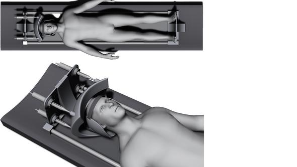
“We have demonstrated that it’s possible to perform a clinical test, called the Spurling test, using a hydraulically controlled device inside the MRI machine on healthy pilot patients and real patients with radiating nerve pain,” says John Hutchins, continuing:
“The protocol includes maneuvering the patient’s head upward and tilting it towards the side where symptoms are present, followed by gentle rotation and the application of a compressive force. Subsequently, MRI images are captured while the patient maintains this induced position. By analyzing these images, we can observe variations in how the nerve exits are impacted, offering a comparative study between the patient's neutral state and the provoked, symptomatic state.”
“A promising advancement”
What significance could this research have for how this patient group is assessed in the future?
“Functional position MR imaging represents a promising advancement in the diagnosis of foraminal stenosis, particularly for patients whose symptoms are dependent on their body’s positioning. Despite facing certain challenges and limitations, our initial findings underscore the substantial benefits and warrant continued investigation and enhancement of these imaging techniques.”
What has been enjoyable, and what has been challenging about the doctoral project?
“It has been incredibly rewarding to develop a hydraulically controlled compression device that works in practice. The challenging part is that everything requires more time and resources than anticipated.”
Text: Jakob Lundberg