
Jens Johansson: New method for 3D imaging with whole brain coverage
He has created a new method for three-dimensional imaging of the entire brain using magnetic resonance imaging. Such 3D measurement could potentially aid in the investigation of small lesions in the brain that may otherwise be difficult to detect. This is shown by Jens Johansson in his doctoral thesis.
JENS JOHANSSON
Dissertation Defense: December 1, 2023 (click for details)
Doctoral Thesis: Diffusion MRI: aspects of reproducibility and novel segmented 2D and 3D approaches for higher resolution and geometric fidelity
Research Area: Radiology and Imaging
Sahlgrenska Academy, The Institute of Clinical Sciences

Jens Johansson is a medical physicist at Sahlgrenska University Hospital and is working on trying to improve image quality in magnetic resonance imaging (MRI) examinations.
He focuses on a special type of MRI technique called diffusion-weighted imaging (DWI). This technique measures the random movement of water molecules, known as diffusion.
“The DWI-technique is a very useful tool for non-invasive imaging and clinical investigation of the human body, for example to localize and characterize lesions. However, the technique suffers from several inherent limitations, which prevent it to reach its full clinical potential,” says Jens Johansson, who lists a few examples:
“Low spatial resolution, image distortions, image blurring, and motion-artifacts.”
Increased resolution and signal
Jens Johansson’s research aims to investigate these limitations associated with DWI and develop novel methods to reduce them.
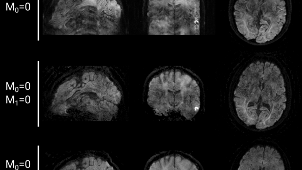
“For example, we have developed a method for 3D-DWI measurement of the entire brain. While DWI predominantly relies on a 2D acquisition scheme, a 3D imaging approach allows for high isotropic spatial resolution at superior signal-to-noise ratio compared to its 2D counterpart. Previously 3D-DWI approaches collect the signal from several thin slabs of the brain, which is later combined during image reconstruction. Our new 3D-DWI method instead acquires the data using a single thick-slab with full brain coverage.”
What are the main findings of the thesis?
“The thesis concludes that single thick-slab 3D-DWI with full brain coverage can be made possible with today’s modern MRI cameras.”
“Capture early changes”
What patient benefits could your research findings provide in the future?
“It could help to find small changes that are otherwise not visible. A 3D-DWI measurement can also provide multi-planar reconstruction, which means that you can rotate the image and check at any angle,” says Jens Johansson, and continues:
“The 3D-DWI approach can potentially aid in the diagnosis of small brain lesions. And the multi-planar reconstruction of images in different planes could help doctors get a better understanding of how, for example, a tumor grows in three dimensions.”
Jens Johansson points out that further research is needed to establish the 3D-DWI as a robust imaging alternative.
“For example, the acquisition time for 3D-DWI needs to be shortened and the method needs to be evaluated in patients. This thesis demonstrates that our new method is a viable venue for single thick-slab 3D-DWI with full brain coverage.”
Text: Jakob Lundberg