
Correlative Array Tomography
Correlative Array Tomography combines different microscopy modalities to explore the 3D cellular architecture of large samples in extremely fine structural and molecular detail by, for example, merging the molecular discrimination of light microscopy with the ultrastructural imaging of electron microscopy.
SciLifeLab Unit in Correlative Array Tomography since 2021
We are part of the Integrated Microscopy Technologies infrastructure which also includes the Advanced Light Microscopy and Focused Ion Beam SEM units.
Correlative multimodal imaging
CCI specialises in three-dimensional correlative multimodal imaging (CMI), combining multiple imaging techniques to generate comprehensive views of biological specimens. By integrating complementary information across spatial scales from nanometres to millimetres, CMI provides insights into structure, function, dynamics, and molecular composition that cannot be achieved with a single imaging modality alone.
Correlative array tomography
Array tomography (AT) combines light and electron microscopy to enable high-resolution 3D imaging of large biological samples. Fluorescence AT provides enhanced resolution and molecular multiplexing, while electron AT offers efficient ultrastructural reconstruction. Correlative Array Tomography (CAT) integrates both approaches, combining molecular information from fluorescence microscopy with ultrastructural data from electron microscopy to generate comprehensive and complementary insights into biological systems.
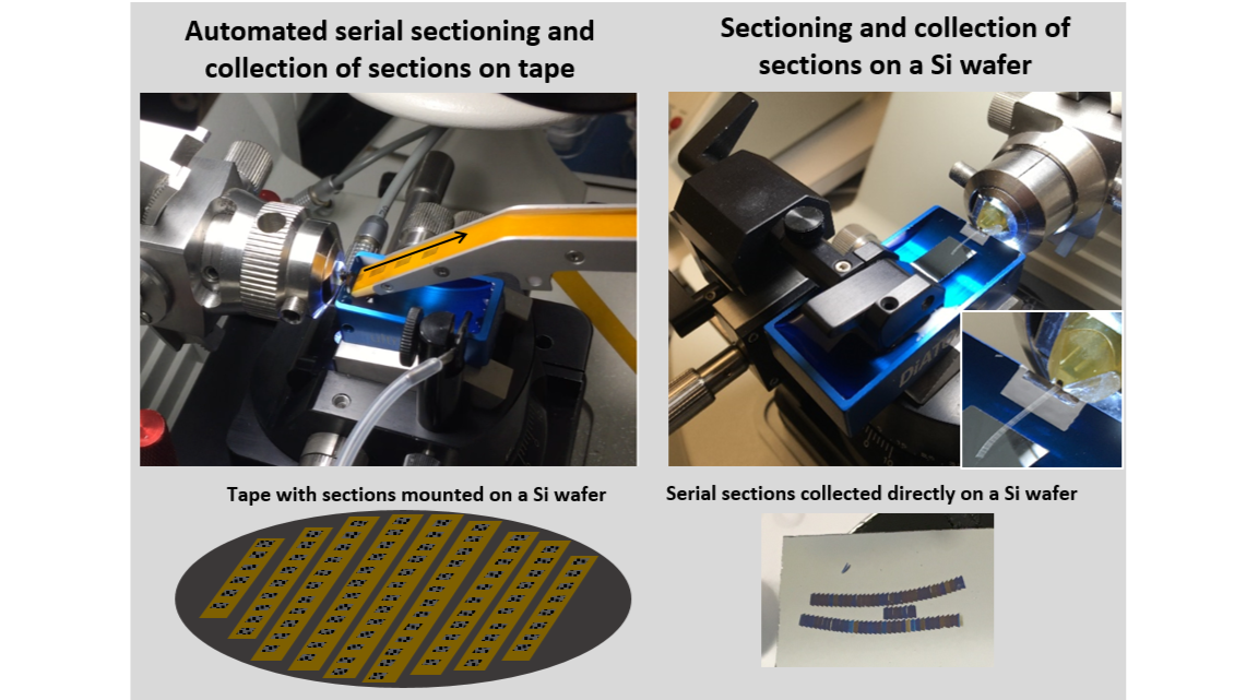
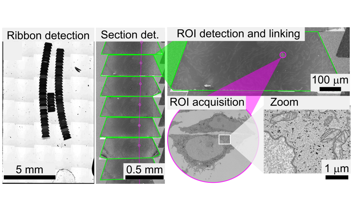
CAT workflow
Unique advantages with CAT
- CAT enables robust registration between the different microscopy modalities, because the same array of ultrathin sections is imaged in the different microscopes.
- CAT, as a non-destructive system compare to en-bloc techniques (FIB-SEM and SB-SEM), permits a certain level of flexibility in experimental design. Additional rounds of immunostaining can be added. Subsequently, upon completion of the electron microscopic imaging, the samples can be revisited again at a later time and reimaged at the SEM at a different magnification, or in a different region. The image stacks on interest can be examined with new questions in mind.
- When targeting rare or specific events within large populations or tissues, CAT is increasingly being recognized as “the method of choice” due to its ability to cover large sample areas (mm-cm).
Video gallery