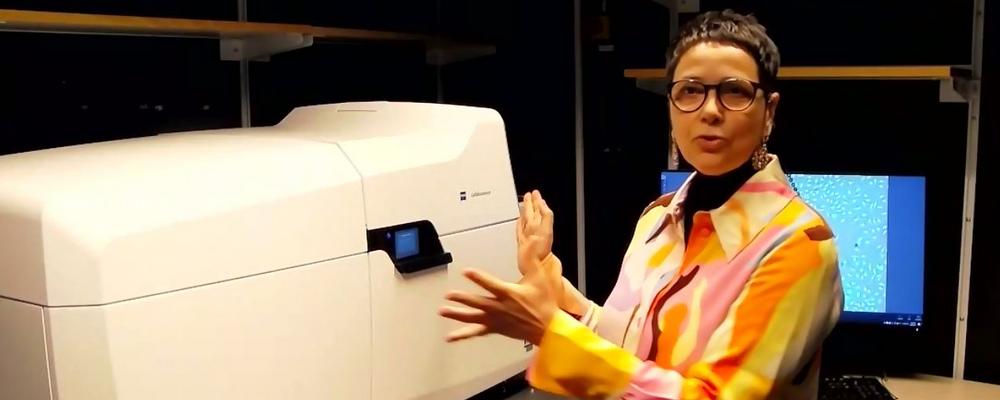
High content images of live cells
A unique feature is that the microscope can combine different light microscopy methods in one single system to create sharp and functional images for specific purposes.
The microscope can, for example, use laser scanning microscopy to construct high resolution three-dimensional images of living biological samples, but can also combine this with large overview images from a sensitive widefield camera. Both the acquisition of such images and their analysis can be fully automated.
Expertise from around the world
“Our staff has cutting-edge expertise in the field and can handle these advanced functions” said Julia Fernandez-Rodriguez, Head of unit at CCI, where researchers from various universities and from industry can turn to for support and training in addition to access various advanced light and electron microscopes.
Julia also explained that the microscope is not just programmable and automated but can also be handled remotely via the user’s own computer.
Jens Wigenius, from the manufacturer Zeiss, also emphasizes that it is in combination with unique scientific competence that the advanced system can reach its full potential.
“What makes it unique world-wide is the competence at CCI together with the possibilities that the microscope offers. Researchers will be able to see details that they have not previously been able to see, and also be able to collect a larger amount of data from the samples,” Jens Wigenius said.
“I am really curious about which projects the team and the researchers at the university will be able to carry out with this instrument in the future.”
Technology as catalyst
Both Julia and Jens participated in an online opening of the microscope that gathered almost 60 people via Zoom. On video link from Jena, Germany, Sören Prague, application specialist at Zeiss, also participated. He gave a demonstration of some of the many functions and highlighted the role of technology in research.
“I hope that new ideas and projects can be developed and implemented with the help of the technical possibilities that come with the Celldiscoverer 7. Sometimes it is new technology that can enrich and push the development within research, and with this microscope only creativity sets the limits” Sören Prague said.
Features beyond the ordinary
Through this instrument, you can both get a full-scale overview of your samples but also zoom in on details at the 150nm scales, thanks to the Airyscan confocal system. You can also manage, edit, and work in 2D and 3D views.
Another important feature is that the microscope is stable in temperatures as varied as from 14 to over 40 degrees Celsius. Samples can be analyzed in different environments, and automatically change from dry to water immersion objectives. The lenses are protected inside the microscope, which makes the instrument less fragile, more durable, and safe for remote operation.

