Specimen Preparation for LM
In the light microscope many different types of specimens can be imaged, These includes live or fixed cells and tissue and there are many ways to prepare these specimens.
There are many ways to prepare specimens for light microscopy. Generally, our users perform the sample preparations in their own lab and come with the prepared sample to the microscope. If needed, we can give advice about fixation protocols, fluorophore selections, mounting media etc.
For big tissue samples we can perform different tissue clearing protocols as a service. In addition, there is a cryostat at the CCI facility, which can be booked by the hour like our other equipment.
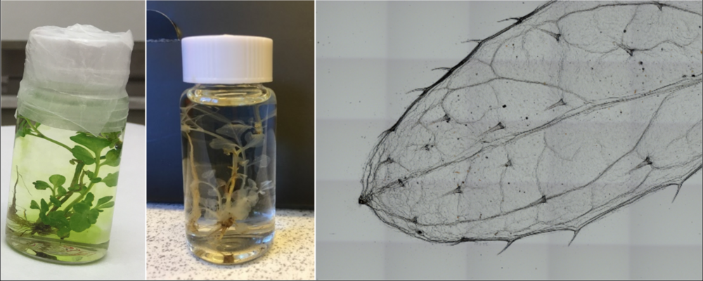
The tissue clearing protocols aim to make tissue as transparent as possible to enable much greater imaging depths. Using this technology, researchers can investigate entire structure of tissue without sample sectioning, for example, an entire 3D map of neuronal pathways can be imaged from a cleared mice brain.
CCI provides tissue clearing services to researchers. Please contact us, preferably using cci@gu.se, if you are interested in tissue clearing technology, and we can find an ideal clearing protocol for your sample and your research goal using our modified tissue clearing protocols.
Full service includes:
- We design and provide an ideal clearing protocols for your sample.
- We will then take care of the technical side of tissue clearing.
- This service is subject to availability of our resources.
We currently offer the following protocols:
- Advanced CUBIC
- ClearSee
- CLARITY
