Olympus cell^R and scan^R - screening station
This equipment combines the scan^R screening platform and cell^R imaging station in one system, which provides unmatched flexibility for both routine and advanced applications of fluorescence microscopy. It is integrated with a live cell incubation system for long time lapse studies.
The scan^R is an imaging platform, built on an inverted IX81 motorized microscope, designed for fully automated image acquisition and data analysis of biological samples. It is especially suited for high content screening/analysis of samples in multi-well plates, but almost any other format is also possible to use e.g. single slides, Petri dishes or custom-built arrays. There is a second license of the analysis software installed on a powerful analysis computer.
The cell^R part, where advanced protocols can be set up in the xcellence software, is another option for live cell imaging as well as for imaging of fixed materials.
This system is equipped with a Xe light source (MT20), a Hamamatsu C8484 CCD camera, hardware and software autofocus, an automatic stage, and an incubator for live-cell imaging.
Objectives
There are several types of objectives available, both long-distance objectives, suitable for ordinary multi-well plates with thick plastic bottom, and high-resolution objectives for thin carriers:
UPLSAPO 4x/0.16
UPLAFL 10x/0.30 Ph1
UPLSAPO 20x/0.75
UPLSAPO 40x/0.95
LUCPLFLN 20x/0.45 Ph1
LUCPLFLN 40x/0.6 Ph2
UAPO 40x/1.15 W
Filter options
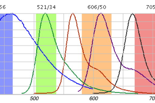
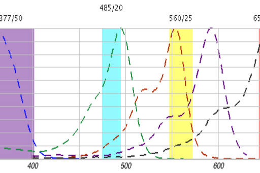
To facilitate fast switching between fluorofore channels there is a quadrupel emission filter set, see figure 1, and a fast rotating filter wheel with excitation filters, see figure 2. In addition to these filter combinations there are also filters available for CFP/YFP and FURA2.