
Jasmine Bagge: Optimized preservation reduces tissue injury prior to intestinal transplantation
The research presented in Jasmine Bagge’s doctoral thesis explores how preservation of the small intestine can be optimized prior to intestinal transplantation. The results show that filling the intestine with a PEG-based solution can reduce damage to intestinal tissue.
JASMINE BAGGE
Dissertation defense: 22 January 2026 (click for details)
Doctoral thesis: Mechanisms of mucosal injury and repair in intestinal transplantation
Research area: Surgery
Sahlgrenska Academy, The Institute of Clinical Sciences
Intestinal transplantation is a complex and relatively rare procedure associated with high risk of serious complications, infections, and organ rejection. The surgery is mainly performed in patients with intestinal failure who develop complications following long-term treatment with intravenous fluids and nutrition.
“One contributing factor to early complications is the injury that occurs during the time when the intestine is removed from the donor and before it is transplanted into the recipient—during this period the intestine lacks blood supply and is affected by oxygen deprivation. During this time, the intestine is stored on ice in a preservation solution before transplantation,” says Jasmine Bagge, resident physician in pediatrics at Queen Silvia’s Children’s Hospital and a doctoral student at the Institute of Clinical Sciences.
When blood flow is later restored to the organ during transplantation, the intestine undergoes further deterioration known as reperfusion injury. This may lead to additional tissue damage and increase the risk of infection.
PEG-based solution as a complement to currently used intestinal preservation
“Our research group has focused on different methods to optimize preservation of the small intestine prior to transplantation. Among other things, we have studied the intestinal mucosa and investigated whether a polyethylene glycol (PEG)-based solution, used in combination with currently available preservation solutions, can better protect the intestine while awaiting transplantation and reduce tissue damage.”
The results show that preservation using a PEG-based solution has a protective effect on the intestine. This could potentially extend the time the intestine can be kept on ice prior to transplantation and reduce the risk of post-transplant complications.
“We also studied the effect of continuous air-insufflation to see whether this could further optimize and prolong preservation time. However, the results did not show any major advantage compared with filling the intestine with the PEG solution alone.”
Additionally, when the colon was studied, the researchers noted that the colonic mucosa was well preserved for at least eight hours of cold preservation, and after 24 hours only minor injuries were observed.
What has been the most rewarding and challenging part of your doctoral project?
“It has been rewarding to conduct preclinical research in parallel with clinical work. I have also had an excellent supervisor and co-supervisor who supported me throughout the years. What has been challenging, but also very rewarding, has been learning new laboratory methods.”
Text: Susanne Lj Westergren
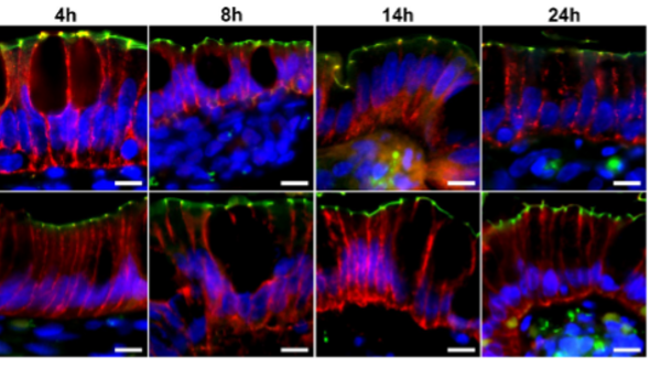
Illustration from thesis: Immunofluorescence microphotographs showing the tight junction protein ZO-1 (green) expressed along the apical membrane and claudin-3 (red) found on the basolateral membrane in the control group (vascular perfusion, VP) and in the intervention group (vascular preservation and luminal perfusion, VP-LP). Nuclei were stained blue using 40,6-diamidino-2-phenylindole. Original magnification x400, scale bar 20 mm.