Newly appointed professor, head of research area, researcher, academic representative, and society chair. Radiologist Åse Johnsson holds many roles — but at the center of them all is the medical image: how it is captured, reviewed, interpreted, and taught.
We meet in a dark room in the radiology department. On one of the screens, Åse Johnsson displays a CT scan produced with conventional technology. On the other: the same patient, imaged with the new photon-counting CT system.
The difference is striking.
“The new technology offers much better contrast, less noise, and clearer detail. You get high-resolution images in several planes — and an important advantage is that it can be done with a lower radiation dose than previous generations of CT.”
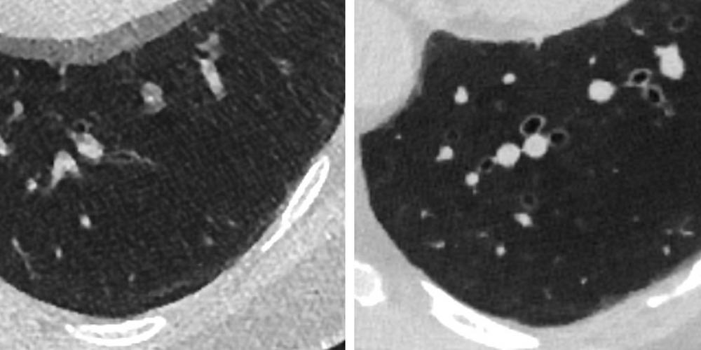
Image
CT images of the same lung using two generations of CT technology. Left: energy-integrating CT, largely unchanged for about 50 years. Right: photon-counting CT, introduced only a few years ago.
Most spots are completely harmless, but a small proportion can be early signs of lung cancer
She rotates the image, changes perspective, zooms in. Small dense areas in the lung become visible.
“Most of these spots are harmless, but a small proportion can be early signs of lung cancer. One of the major challenges is identifying which spots actually need follow-up,” says Åse Johnsson, professor and head of research area for radiology and imaging at the Institute of Clinical Sciences.
Professor since October
After eleven years as senior lecturer, Åse Johnsson became professor of radiology and imaging in October.
“It is incredibly rewarding to contribute to the field at a new level and become part of the national community of professors in radiology,” she says.
She also works clinically as a thoracic radiologist and plays a central role in SCAPIS, Sweden’s largest population-based study focusing on the heart and lungs.
In the ongoing SCAPIS follow-up, 15,000 participants are being examined using the new technology that provides sharper images.
“The lungs are highly sensitive to radiation. When many exposures are involved, the possibility of reducing dose over time becomes very important,” she says.
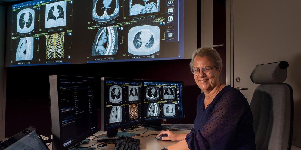
Image
In a dark room at the radiology department, Åse Johnsson reviews CT images and teaches medical students.
Photo: Jakob Lundberg
Research, teaching, and clinical work
What drives you in all this? “I’m passionate about research, about supporting students, and about the organizational perspective — not only within radiology but also to help ensure that the new medical program as a whole becomes as strong as possible.”
Åse Johnsson has been course director for semester 7 and is now planning semester 11 in the new program. She teaches medical students in six of the program’s twelve semesters, leads specialist-training courses, and supervises those who teach radiology.
“I meet every student who chooses to take our courses. It’s a great responsibility but also a privilege. Radiology is a piece of the puzzle in almost every part of healthcare.”
AI soon in clinical routine
She works both at the university and clinically at Sahlgrenska University Hospital. Her research includes low-dose imaging techniques and how AI can detect subtle changes in medical images. “We use AI to identify nodules — the small spots in the lungs — and we’ve done so in SCAPIS for several years. I’ve advocated for ten years to bring AI into clinical routine, and now we’re closer than ever.”
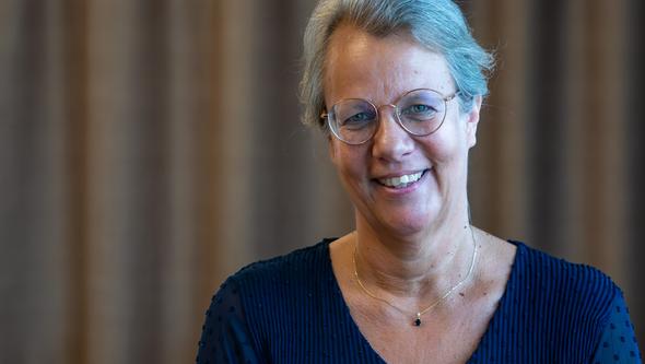
Åse Johnsson, professor and head of research area in radiology and imaging, works in three fields: clinical radiology, medical education at the University of Gothenburg including curriculum development and teaching, and research.
Photo: Jakob Lundberg
MRI — like a pipe organ of radio waves
Åse Johnsson’s deepest technical interest lies in magnetic resonance imaging, especially cardiac MRI. “It’s absolutely fascinating to image a beating heart. We can measure motion, flow, and tissue changes — all without radiation. It’s like an pipe organ made of radio waves. And the development will continue in many ways.”
It’s absolutely fascinating to image a beating heart
She began her research career with an experimental thesis on titanium implants in bone tissue before shifting her focus to thoracic radiology. “I wanted my research to connect with my clinical work — and now it does.”
A fluid line between work and free time
Åse Johnsson is also the academic representative in the management group for radiology and chair of the Swedish Society of Thoracic Radiology.
“Like so many others in academia, I work a great deal, but the line between work and free time is sometimes fluid. Radiology is not just a job for me — it is also a passion. It’s absolutely worth all the time I put into it. Everything connects — research, education, and the profession. I want to help move things in the right direction.”
Energy-integrating CT (EID-CT) – Clinical standard in Sweden since the mid-1970s – Sums the total energy of all X-ray photons – Technique has improved, but the principle is unchanged
Photon-counting CT (PCD-CT) – Introduced clinically in Sweden in 2022–2023 – Counts individual photons and measures their energy – Provides higher resolution and less noise – Enables lower dose, depending on protocol and clinical need