Fetal cardiac intervention may reduce risk of single-ventricle defect
Can a catheter-based intervention during fetal life increase the likelihood that children with a narrow heart valve are born with two functioning ventricles? Mats Mellander, adjunct professor of pediatrics at the Institute of Clinical Sciences, is one of the leading researchers in an international study examining whether balloon dilation can be an effective treatment for certain fetuses.

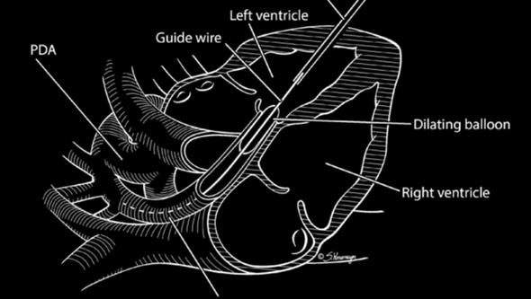
In hypoplastic left heart syndrome, HLHS, the left ventricle is so underdeveloped that it does not contribute to the circulation. This heart defect can arise when the aortic valve is narrowed during fetal life, a condition called fetal aortic stenosis.
The left ventricle may then develop so poorly that the child is born with a single-ventricle heart, where survival after surgery in the neonatal period is lower than when the heart has two well-developed pumping chambers.
Observational study since 2021
Since 2021, an international prospective observational cohort study has been ongoing on balloon dilation of the narrowed valve during fetal life, with researchers in Gothenburg holding primary responsibility.
Outcomes in fetuses who have undergone the intervention are compared with those in a control group in which the pregnant women have chosen to refrain from treatment.
“The woman makes the decision in consultation with her physician. It is always made entirely independently of the study, which does not influence clinical management either,” says Mats Mellander.
Once the decision has been made, participation is offered regardless of whether the woman has accepted or declined the intervention. So far, around 100 fetuses have been included, and the goal is 150.
“We want to examine whether the intervention can promote growth of the left ventricle so that the child is born with two functioning cardiac ventricles. That would be a major advantage for the child.”
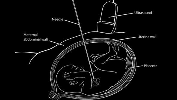
Intervention at 23–32 weeks of pregnancy
The cardiac intervention is performed between gestational weeks 23 and 32 under general anesthesia. Swedish patients who wish to undergo the treatment are referred to Linz, Austria, which has the greatest experience with this procedure in Europe.
The study is conducted in close collaboration with Annika Öhman, pediatric cardiologist at the Pediatric Heart Center at Sahlgrenska University Hospital.
“We do not yet have robust scientific evidence that prognosis improves. Published case series suggest improvement in selected cases, but no prospective study with a comparison group has previously been conducted,” says Mats Mellander.
Risks and uncertainty
Balloon dilation carries a five to ten percent risk of fetal death.
How do you help expectant parents make a decision when definitive evidence is lacking and there is also a risk associated with the intervention?
“We provide information about the potential benefit for the child and the risk to the fetus. At the same time, we are clear that solid evidence is lacking and that the study is intended to provide better guidance for future decisions.”
Fifteen centers in Europe as well as five in the United States and Canada are participating. Coded clinical data are registered in Gothenburg, and ultrasound images are analyzed at a Core Lab by Carina Olausson, biomedical scientist at the Pediatric Heart Center. Data collection will continue until 2028, and the results are expected to be presented thereafter.
Text: Jakob Lundberg