
Jonathan Arvidsson: Development of perfusion MRI techniques
Magnetic resonance imaging (MRI) has a wide range of diagnostic applications. Jonathan Arvidsson’s thesis focuses on MRI techniques for measuring blood flow. The primary goals are to improve the techniques and to explore their diagnostic value in relevant patient groups.
JONATHAN ARVIDSSON
Doctoral Thesis: Advancements in DSC and BOLD perfusion imaging: Acquisition, analysis and clinical application
Research Area: Medical Radiation Sciences
Sahlgrenska Academy, The Institute of Clinical Sciences
A magnetic resonance imaging system (MRI) can provide detailed images of anatomical structures. It can also be made to sensitize various tissue characteristics, making it possible to customize images for specific diagnostic inquiries.
Perfusion refers to the flow of blood through the capillary bed of a tissue. Jonathan Arvidsson’s thesis addresses both the development of MRI techniques for measuring blood flow as well as the application of these techniques in the brain and peripheral calf muscle.
Two different imaging techniques
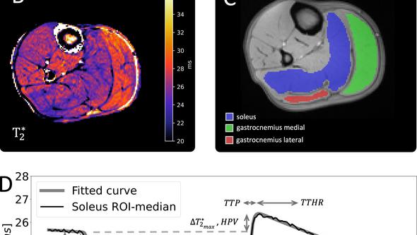
The first part of the thesis is focused toward perfusion imaging techniques where blood flow contrast is enhanced by an intravenously administered contrast agent. The second part is based on blood oxygen level-dependant magnetic resonance imaging, which leverages signal properties of venous blood. The signal effect in this latter technique is enhanced by temporarily limiting the blood flow to the calf muscle by use of a pressure cuff.
“Two subprojects have focused on technical considerations. For instance, determining how the duration of contrast agent injection in DSC-MRI and the significance of the cuffing duration in cuff-based BOLD-MRI affects the blood flow measurements,” explains Jonathan Arvidsson, a biomedical engineer working at the department of biomedical physics and engineering at Sahlgrenska University Hospital.
Two patient groups studied
His clinical work centers around two medical conditions: idiopathic normal pressure hydrocephalus, a disease characterized by increased volume of the brain ventricles with neurological and cognitive disturbances being part of the disease symptoms – and peripheral artery disease, in which artery narrowing or blockage causes reduced blood supply to the legs and symptoms of walking pain and in severe cases impaired wound healing.
What are the most significant research findings?
“I think our results with the BOLD-technique has shown the techniques sensitivity. In part sensitivity to the cuffing duration, but also in its ability to separate between patients with peripheral artery disease and healthy control subjects.”
The long-term objective
What practical benefit can this research offer?
“A long-term goal for both patient groups we are working with is for MRI techniques to aid in identifying which individuals may benefit from surgery,” Jonathan Arvidsson says.
What has been enjoyable and challenging about this doctoral project?
“For me, there’s joy in collaborating with colleagues who share a passion for magnetic resonance imaging and research. My project has taken unexpected turns, incorporating two different MRI techniques and two distinct patient groups. I’m very pleased to have finalized it in this way.”
Text: Jakob Lundberg
