
Chiara Micheletti - Multimodal and multiscale characterization of bone and bone interfaces in health and disease
In June 2023 Chiara Micheletti defended her thesis for Doctor of Philosophy in Medical Science at the Institute of Clinical Sciences, Sahlgrenska Academy, in the research subject of Biomaterial science.
The title of the thesis: Multimodal and multiscale characterization of bone and bone interfaces in health and disease
Link directly to the doctoral thesis
Multimodal and multiscale characterization of bone and bone interfaces in health and disease
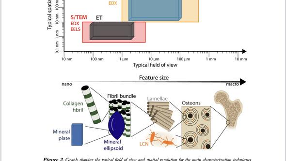
Our understanding of phenomena often involves their direct observation. However, bone architecture is challenging to visualize given its multi-level hierarchical organization. In this thesis, bone and bone interfaces are characterized via multimodal and multiscale platforms, combining different techniques across several length scales.
“Bone is a hierarchical material, meaning that smaller components are progressively organized into larger structures. This multi-level architecture starts at the nanoscale with collagen fibrils and a calcium phosphate-based mineral and culminates at the whole bone level where we can distinguish between cortical and trabecular bone , ” says Chiara Micheletti.
She is a PhD student completing a double PhD degree (cotutelle) between the Department of Biomaterials at the Sahlgrenska Academy and the Department of Materials Science and Engineering at McMaster University (ON, Canada).
Her PhD research took place in between these two institutions, where she focused on examining bone and bone interfaces with advanced characterization tools, including but not limited to electron microscopy and spectroscopy.
“Seeing is believing”
The complex organization of bone is hard to visualize because different techniques are required to obtain information about different length scales. However, as the proverb says, “seeing is believing”, and our understanding of things is often better accomplished by looking at images.
“Therefore, in my research, I apply a characterization approach more typical of materials science to visualize bone and bone interfaces across multiple length scales to obtain information about structure and composition. This characterization approach is multiscale because it targets different length scales in bone, and multimodal because it is accomplished by different techniques.”
Focus on bone in both healthy and diseased conditions
The main tools used in this research are micro-computed X-ray tomography, electron microscopy and spectroscopy (both scanning and transmission electron microscopy), and Raman spectroscopy. These are applied to both human and animal bone samples “ex vivo”, namely retrieved from patients or from animal studies.
“My PhD thesis focuses on bone in both healthy and diseased conditions such as type 2 diabetes, osteoporosis, and medication-related osteonecrosis of the jaw. The main objective of my doctoral research is to understand the effect of disease-induced changes on bone structure and composition, as well as on bone repair in the presence of biomaterials.”
This knowledge can be used to identify new bone implant solutions that, for example, reduce fracture risk by improving bone regeneration in aging-related bone diseases such as osteoporosis. These potential applications are becoming increasingly relevant especially as the world population ages. Lastly, there is still a debate on how healthy bone organizes itself at a fundamental level, so my research also focuses on understanding its architecture at the nanoscale. This is a critical aspect to understand, as bone structure is built from the bottom-up.
The importance of three-dimensional imaging with nanometer resolution
“By using a multimodal and multiscale characterization approach, my doctoral research enabled us to obtain relevant information about the structure, composition, and repair of bone, which are especially relevant to understand disease-induced changes and abnormalities. These findings will contribute to the development of bone implants.”
Lastly, advanced three-dimensional imaging at the nanoscale was able to answer some long-debated aspects regarding bone’s smallest components (collagen fibril and mineral crystals). In the future, these tools can be applied to bone in compromised conditions to better understand how diseases affect bone structure and composition starting from its building block units.
Editor: Susanne Lj Westergren
Supervisor: Anders Palmquist
Co-supervisor: Kathryn Grandfield and Furqan A. Shah
Opponent: Håvard J Haugen, Institute of Clinical Dentistry, University of Oslo, Oslo. Norway
Examination board: Klara Sjögren, Michael Noseworthy, Joey Kish, Fang Liu and Anders Björkman