
Biomineralization and Biointerfaces
Short description
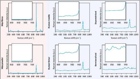
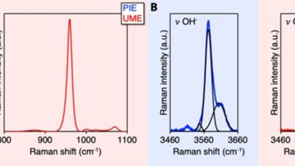
The Biomineralization and Biointerfaces Group investigates “bone as a material”, examining the structure, composition, and adaptation under diverse conditions using advanced techniques such as electron microscopy, micro-Raman spectroscopy, and X-ray micro-computed tomography, encompassing the macro-, micro-, and nanoscale. Bone is a complex, living tissue with a hierarchical architecture, from tiny mineral crystals to the entire skeleton. Particular attention is on the mineral component of the extracellular matrix and the mineralisation process of osteocytes (micropetrosis), bone repair biomaterials such as calcium phosphates, titanium, magnesium, and cobalt-chromium alloys, and distinguishing bone mineral from other bioapatites (e.g., in dental enamel) and geological apatites (i.e., hydroxy(l)apatite).
About our research
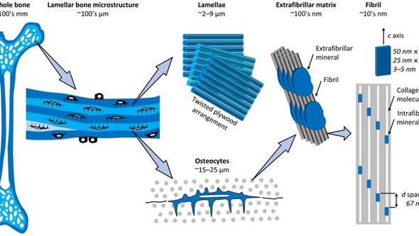
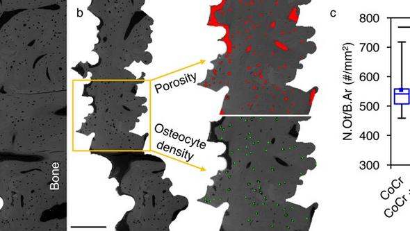
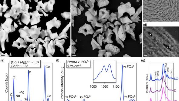
Our group investigates mineralised tissues, particularly bone, from a materials science perspective, focusing on structure–property–function relationships under diverse conditions. This is achieved using advanced analytical techniques, including scanning electron microscopy (SEM), transmission electron microscopy (TEM), energy dispersive X-ray spectroscopy (EDX), micro-Raman spectroscopy, and X-ray micro-computed tomography (micro-CT), enabling analysis of material properties (i.e., structure and composition) across macro-, micro-, and nanoscale levels. Bone consists of either a porous trabecular framework or a dense cortical structure, both forming lamellar bone (Figure 1). The twisted plywood arrangement of lamellae results from alternating fibril orientations. Osteocytes, residing in lacunae interconnected by canaliculi, regulate bone remodelling. Type-I collagen molecules and carbonated apatite crystallites form a nanocomposite structure within collagen fibrils.

GROUP MEMBERS
Doctoral students
Current:
Josefin Ewerman
Magdalena Korytowska (Malmö University)
Previous:
Martina Jolic
Heithem Ben Amara
Chiara Micheletti (McMaster University, cotutelle)
Krisztina Ruscsák
Master’s thesis students
Previous:
Ellinor Klippmark (Karolinska Institutet)
Emily Petterson (Chalmers University of Technology)
Märta-Sofie Geijer
Undergraduate students
Current:
Mohammad Amairi
Nick Kavoszade
Yasmin Sinan Sami**
Rahaf Ismaeil
Previous:
Mariana Cavero Rozas#
Hanna Gundewall**
Khaled Rslan**
Fabiana Benedini Galli Zambardino#
David Eskandar-Baghbani
Adyan Aziz**
Martina Ariana Ghoraishi**
Isabella Åberg*
Zeinab Jalil**
Sonali Sharma
Annika Juhlin
Samad Mirzayev
Edvin Jergéus
Supported by:
*Axel Lennart Larssons fond
**Stiftelsen Mary von Sydows, född Wijk, donationsfond
#International Federation of Medical Students’ Associations
