
Tracking changes in the gut’s protective barrier in real time
From a passive layer of mucus to a sophisticated defense system against the gut’s bacteria. Research at the University of Gothenburg has changed our understanding of how the gut protects itself. Now Malin Johansson and her colleagues will be able to track rapid processes in the mucus at higher resolution than before.
A SEK 4.8 million grant from the IngaBritt and Arne Lundberg Research Foundation will allow researchers at Sahlgrenska Academy to add a new high-speed, high-resolution microscope to their already advanced microscopy capabilities.
For Malin Johansson, Professor of Biomedical Laboratory Science specializing in Biochemistry at the University of Gothenburg, the new instrument opens up new possibilities.
“We’ll be able to resolve both time and space in a completely different way. We can see where something happens, when it happens, and what happens next. These are questions we’ve wanted to tackle for a long time. It’s fantastic to have this opportunity, and we’re very grateful,” says Malin Johansson.
Different ways of viewing the same tissue
The researchers already use several forms of advanced microscopy. One of these methods is electron microscopy, that allows them to examine extremely small structures in detail, but the tissue must be fixed or frozen. The new microscope adds something different: high resolution combined with rapid imaging of live samples.
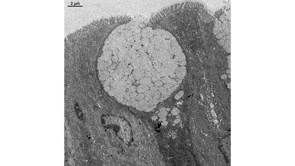
This is particularly valuable when studying mucus, which consists largely of water and can change during sample preparation.
When the protective barrier fails
One area in which Malin Johansson plans to use the new technology is ulcerative colitis, where the gut’s protective mucus barrier is altered. Her group also studies what happens as long-term inflammation progresses toward colorectal cancer.
Malin Johansson also studies Environmental Enteric Dysfunction (EED) together with Associate Professor Thaher Pelaseyed. The condition primarily affects children in areas where malnutrition and infections are common and can impair nutrient absorption and growth. The project is supported by the Gates Foundation.
![[none]](/sites/default/files/styles/50_10_6_xmedium_1x/public/2026-09/Malin%20Johansson%20Sept%202026%2016x6.jpg?h=025155af&itok=0Le7ptiL)
“We still know far too little about why EED develops. If we can understand what happens to the mucus barrier, we may be able to identify markers of the disease and, in the longer term, find ways to treat it.”
A strong research community
Malin Johansson’s group is part of the Mucin Biology Groups, where researchers with different areas of expertise work closely together. Their work spans fundamental biochemistry and structural biology, cell biology, advanced microscopy, and research using patient samples.
The research also extends beyond the gut. Researchers in this community have helped reshape our understanding of mucus in the airways and also study mucus in the female reproductive tract.
“None of us can do all of this alone. But together, we bring a very broad range of expertise, from molecules to patient samples. When we bring all of that expertise together, we can go much further and ask questions we otherwise wouldn’t be able to ask. It’s very inspiring,” says Malin Johansson.
