
Thyroid development mapped during fetal growth
Researchers have mapped how the two distinct endocrine cell lineages of the thyroid gland develop separately before later merging into a single organ. The study provides new insight on how the thyroid forms in mammals and also shows that similar biological processes can be dysregulated in aggressive thyroid cancer.
The study is published in Nature Communications and is the result of a collaboration between researchers at the University of Gothenburg and UMass Chan Medical School together with collaborators in Italy.
The thyroid gland consists of two types of hormone-producing cells that regulate the body’s metabolism and calcium balance. In mammals, these cell types merge into a single organ, whereas in other vertebrates they develop into separate organs. How this fusion occurs has long remained unclear, even though developmental defects can lead to congenital abnormalities of the thyroid gland.
Links to thyroid cancer
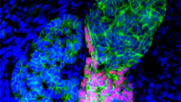
The researchers were now able to follow, step by step, how the two cell types develop, migrate, and integrate into the same organ during embryonic development. By analyzing individual cells, they also identified the genes that govern how cells develop and organize as the thyroid gland forms.
Mikael Nilsson, professor at the University of Gothenburg and one of the study’s senior authors:
“We see clear parallels between how cells behave as the thyroid forms during embryonic development and how certain tumor cells later become invasive. This suggests that cancer cells may reactivate biological programs that are normally active only during organ formation,” he says.
![[none]](/sites/default/files/styles/50_10_6_xmedium_1x/public/2026-06/Sk%C3%A4ldk%C3%B6rteln%20duobild%2016x9.jpg?h=e6d74438&itok=yj-5kRDR)
Cells in development
The study is based on analyses of individual cells and tissues from mice at different stages of embryonic development. The researchers combined developmental biology, tumor biology, and bioinformatics to track how cells change as the thyroid gland forms.
René Maehr, associate professor at UMass Chan Medical School:
“By following cellular differentiation over time, we were able to see how different cell types change, migrate, and gradually integrate into the same organ during embryonic development. This allowed us to gain insight on molecular changes in individual cells to how tissue forms and organizes itself,” he says.
The researchers also found that some of the biological processes involved in thyroid organogenesis during embryonic development are also seen in a rare and aggressive form of thyroid cancer. The findings may therefore contribute to a better understanding of both congenital thyroid abnormalities and how certain forms of thyroid cancer acquire invasive properties.
Article: Resolving thyroid lineage cell trajectories merging into a dual endocrine gland in mammals
