
The placenta provides new clues to impaired fetal growth
Changes in the placenta’s blood vessels may help explain why some fetuses fail to grow as expected during pregnancies affected by the autoimmune disease systemic lupus erythematosus (SLE). At the same time, the researchers did not observe inflammatory changes that have been suspected to play a central role. The findings come from the University of Gothenburg.
Women with SLE (systemic lupus erythematosus) are at increased risk of fetal growth restriction. Although the disease was well controlled and disease activity was low in most of the women included in this study, giving birth to infants small for gestational age remained a common complication.
The study is published in the Journal of Internal Medicine.
Blood supply in focus
The researchers followed 83 pregnancies in women with SLE and 67 pregnancies in healthy pregnant women. Blood samples were collected throughout pregnancy, and after delivery the placentas were examined to identify placental changes associated with fetal growth.
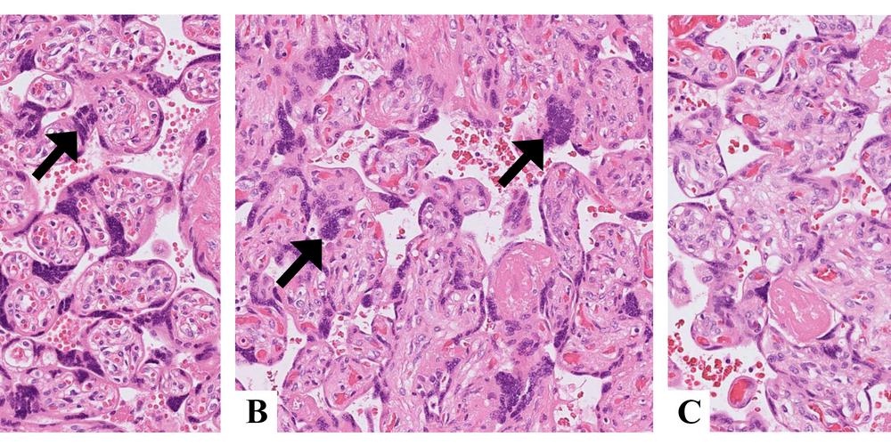
Eight out of ten placentas from women with SLE who gave birth to small-for-gestational-age infants showed signs of impaired placental blood supply. The researchers also identified changes in proteins involved in the formation of blood vessels. In contrast, they did not observe inflammatory changes in the placentas of women whose babies were small for their gestational age.
Marit Stockfelt, researcher at Sahlgrenska Academy, University of Gothenburg, and the study’s lead author:
“Our findings point to the placenta’s blood supply as potentially playing an important role for fetal growth in SLE. This suggests a new hypothesis about the biological mechanisms that may underlie reduced fetal growth in SLE, and helps identify the specific questions future research should focus on.”
![[none]](/sites/default/files/styles/50_10_6_xmedium_1x/public/2026-07/Marit%20Stockfelt%20foto%20G%C3%B6teborgs%20universitet%2016x9_0.jpg?h=a0cf17bb&itok=y54TpgIX)
Toward improved risk assessment
The study identifies associations and cannot determine what causes the observed changes. The researchers also point out that they used small for gestational age as a measure of impaired fetal growth, which is not the same as a clinical diagnosis of fetal growth restriction during pregnancy. In addition, the number of women with SLE included in the study was limited, meaning the findings need to be confirmed in larger studies.
If confirmed, the findings could help identify pregnancies at increased risk of impaired fetal growth. They may also help direct future research toward the biological processes that should be the focus of efforts to develop new diagnostic methods and treatments.
Systemic lupus erythematosus (SLE) is a chronic autoimmune disease in which the immune system attacks the body's own tissues. The disease affects an estimated 44 people per 100,000 worldwide and about 90 percent of those diagnosed are women. It most often develops during the reproductive years.
Many women with SLE can go through pregnancy without serious complications, particularly when the disease is well controlled. However, SLE is associated with an increased risk of adverse pregnancy outcomes, including preeclampsia, preterm birth, and impaired fetal growth.
