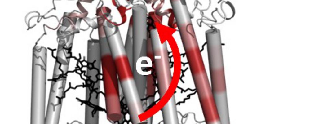
Almost all life on earth has the energy-transducing reactions of photosynthesis as their primary source of energy. These light-driven reactions occur in plants, algae and photosynthetic bacteria.
An X-ray structure of a protein provides scientists with a lot of information as to how they perform their biological task in a living cell.
X-ray films show structural changes within a protein
In this work scientists used a method called time-resolved X-ray crystallography to make a movie of structural changes within the protein responsible for the light-driven chemical reactions of photosynthesis. To achieve this scientists at the University of Gothenburg used a world-leading X-ray source in California (an X-ray free electron laser) to examine if structural rearrangements within photosynthetic proteins occur on the time-it-takes light to cross a hair of your head. Remarkably, these measurements showed that the protein changes structure on this time-scale.
Subtle movements in the protein were seen
Scientists at the University of Gothenburg observed that these movements were very subtle, with both the electron donor (a chemical group which absorbs light and releases an electron) and the electron acceptor (a chemical group which is located 2 nm away and which receives this electron) moving less than 0.03 nm (1 nm = 10-9 m or a millionth of a millimeter) in 300 ps (1 ps = 10-12 sec is called a picosecond and is a millionth of a millionth of a second).
The protein as a whole also changed structure very slightly in order to prevent the electron returning to where it began, which would otherwise make the reaction useless. These results are fundamental to how evolution has optimized energy transducing proteins over billions of years to allow them to perform redox reactions without energy being lost in the process.
– Time-resolved crystallography studies of a photosynthetic protein from bacteria reveal how light-induced electron movements are stabilized by protein structural changes occurring on a time-scale of picoseconds, says Richard Neutze, professor at the University of Gothenburg.
At MAX IV Laboratory in Lund Sweden is building a beamline which will enable scientists to study how protein structural changes occur in other biologically important proteins using the same methods.
