
Magnus Palmér: How imaging can improve the assessment of uterine tumors
Imaging plays an important role in assessing uterine tumors before treatment. Magnus Palmér has compared ultrasound and magnetic resonance imaging (MRI) and explored what advanced MRI techniques can offer.

MAGNUS PALMÉR
Dissertation defense: 27 November 2026 (click for details)
Doctoral thesis: Multiparametric MRI of uterine tumours
Research area: Radiology and Imaging
Sahlgrenska Academy, The Institute of Clinical Sciences
Tumors of the uterine body are common and can be either benign, such as fibroids, or malignant, such as endometrial cancer or sarcoma. Physicians need as much information as possible about these tumors to choose the right treatment.
“Imaging plays an important role in the diagnostic process. Our research has focused on both evaluating and developing preoperative imaging,” says Magnus Palmér, radiologist at Sahlgrenska University Hospital and doctoral student at the Institute of Clinical Sciences.
Ultrasound as a first-line method
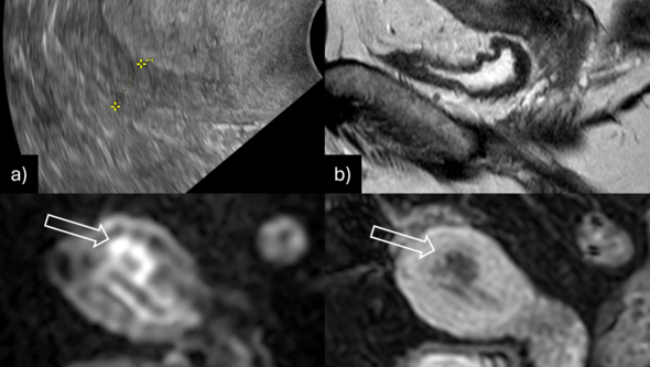
Part of the thesis compares gynecological ultrasound with MRI in endometrial cancer, which affects the lining of the uterus.
“MRI was slightly better than ultrasound at assessing the local extent of the cancer, but ultrasound performed well enough to be used as a first-line method.”
The researchers also investigated how well routine MRI could distinguish between benign and malignant tumors when the diagnosis was uncertain.
“We found that routine MRI was a good second-line method for distinguishing between fibroids and sarcomas in clinically challenging cases.”
Advanced MRI offers more information
Other studies examined whether advanced MRI techniques could provide more information about tumor aggressiveness and possible spread to lymph nodes.
“One advanced MRI technique showed some potential for distinguishing more aggressive tumors from less aggressive ones.”
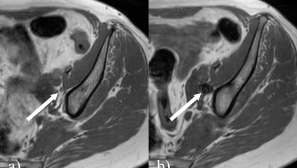
The researchers also used an iron-based tracer injected before the MRI examination.
“This allowed us to localize many lymph nodes relevant to prognosis before surgery, but we could not determine whether they were affected by disease.”
New insights in an evolving field
What has been the most enjoyable part of your doctoral project?
“The most enjoyable part has been everything I have learned, from gaining deeper knowledge of gynecological imaging to learning what it is like to conduct research. I can apply this knowledge both in my clinical work and in future research,” says Magnus Palmér.
And what has been challenging?
“One challenge has been that the field has evolved so rapidly that the role of imaging in the diagnostic process has changed over the course of the project.”
Text: Jakob Lundberg