
Emilia Gryska - AI algorithms – towards a powerful diagnostic tool for brain tumors
On october 7, Emilia Gryska is defending her thesis for Doctor of Medical Science at the Institute of Clinical Sciences, Sahlgrenska Academy, in the research subject of medical radiation science
The title of the thesis is: Automatic tumour segmentation in brain images - moving towards clinical implementation
Link directly to the doctoral thesis

The thesis investigates what is required to clinically implement new artificial intelligence (AI) algorithms for automatic delimitation of brain tumors using magnetic resonance imaging (MRI).
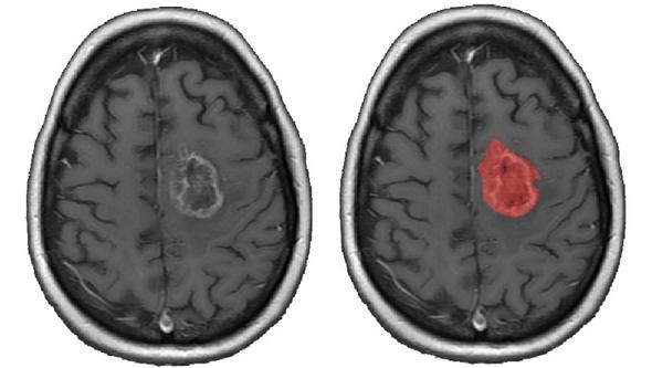
In brain tumors, the volume of the tumor serves as a criterion for assessing its severity. But the volume is rarely measured precisely; it is often estimated visually via MRI images. If the neuroradiologists could instead use AI algorithms that identify and automatically delimit a tumor in an image (segmentation), they would have precise measurements of tumor volume. A segmentation of tumors provides information about the location and volume of the tumor.
The gap between research and application
Even though many studies have developed and validated algorithms for segmentation of brain tumors, it is rarely possible to assess the clinical suitability of the algorithms. They are not described in sufficient detail for the results to be reproducible, something Emilia Gryska now highlights in her thesis. Gryska, who has a background in biomedical engineering, works with medical radiation science at the University of Gothenburg.
She has examined the gap between the research and the clinic with the following three questions:
- Does the research provide evidence that the algorithms will work well in clinical procedures?
- Are radiologists confident AI-generated information, and do they find it helpful?
- What do radiologists need to be able to trust and use the tools?
“In my research work I have long focused on how to implement scientific innovations in clinical practice. I have focused particularly on artificial intelligence algorithms in medical image analysis,” Gryska says.
Reliable AI calculation
To resolve the inability of independent researchers to recreate an algorithm, apply it to the same images, and obtain similar results, she has proposed updated guidelines for designing studies so they can be reproduced.
“I also found that radiologists tend to rely more on the accuracy of the AI-calculated tumor volume, despite a large variation in the perceived segmentation quality, than on their own visual assessment. However, this positive attitude towards AI does not ensure that future clinical tools will actually be trusted and used.”
She also identified several specific trust-related requirements that need to be met to successfully implement AI tools in radiology. The tools, and their introduction into the field, must be reliable and transparent in terms of how they generate the information and how the data are interpreted. In addition, the tools need to be easily implemented and compatible with clinical and day-to-day work.
“The research results partly bridge the gap between the research and the clinic. But future research should focus on testing algorithms in a way that establishes strong evidence for segmentation accuracy in a clinical setting and for how radiologists use the information they receive through the algorithms in their clinical workflow.”
It is also important that the development of AI algorithms provides benefits for patients, in this case faster and more accurate diagnosis.
“Successful implementation of AI tools that increases confidence in the decision support system requires not only collaboration between doctors and tool developers but also understanding and support from hospital management.”
BY: SUSANNE LJ WESTERGREN
Supervisor: Rolf A. Heckemann
Co-Supervisor: Justin Schneiderman och Isabella Björkmann-Burtscher
Opponent: Örjan Smedby, Kungliga Tekniska Högskolan, KTH, Huddinge
Examining Committee: Toshima Parris, Peter Lundberg och Robin Strand