
Kristin Oskarsdotter: 3D-bioprinted tissue could replace lost soft tissue
Using a patient’s own fat cells combined with a supporting biomaterial, new tissue can be 3D-bioprinted in a desired shape. Kristin Oskarsdotter is developing methods that could make it possible to print larger and more stable volumes of fat tissue.
KRISTIN OSKARSDOTTER
Dissertation defense: 23 October 2026 (click for details)
Doctoral thesis: Strategies for promoting the clinical translation of 3D bioprinted adipose tissue
Research area: Plastic Surgery
Sahlgrenska Academy, The Institute of Clinical Sciences
Current methods for reconstructing a breast, for example, often involve harvesting the patient’s own tissue from another part of the body. The amount of available tissue is limited, and the procedure can be extensive for the patient.
“Smaller defects can be treated with injections of the patient’s own fat, but a large proportion of the tissue risks being resorbed over time. This makes long-term outcomes unpredictable,” says Kristin Oskarsdotter, biotechnology engineer and doctoral student at the Institute of Clinical Sciences.
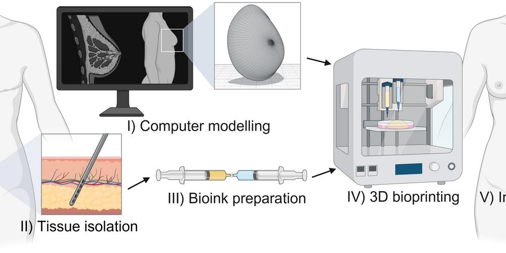
Printing tissue in a desired shape
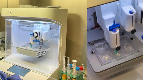
3D bioprinting could offer another way to create the tissue that is needed. The patient’s own fat tissue (adipose tissue) is processed and combined with a supporting biomaterial. The resulting mixture, known as bioink, can then be printed into a predetermined shape.
“My research focuses on developing strategies that promote blood vessel formation and help 3D-bioprinted fat tissue retain its shape over time. Blood supply is crucial for the tissue to survive and limits how much tissue can be printed.”
The ability to retain its shape is also essential if the tissue is to replace lost volume in the long term.
Bioink could improve the tissue
The researchers have investigated how the properties of the bioink can be modified to improve the printed tissue.
“By modifying the supporting biomaterial, we have found that we can influence both the biological activity of the bioink and its stability when 3D-printed implants are implanted in animals.”
The next step is to begin evaluating 3D-bioprinted fat tissue in humans.
“In the future, we hope this could make it possible to create larger 3D-bioprinted tissues and thereby address the substantial clinical need.”
Between technology and clinical care
What have been the most enjoyable and the most challenging parts of your doctoral project?
“As an engineer, navigating the intersection of science and clinical practice has been both challenging and very rewarding. We approach problems somewhat differently. Physicians start with the clinical need and the patient, while engineers tend to focus more on technical solutions,” says Kristin Oskarsdotter and continues:
“Keeping both perspectives in mind has been a valuable learning experience, and it is fantastic to see technological advances actually address a real clinical need.”
Text: Jakob Lundberg