
Correlative Array Tomography
Correlative Array Tomography combines different microscopy modalities to explore the 3D cellular architecture of large samples in extremely fine structural and molecular detail by, for example, merging the molecular discrimination of light microscopy with the ultrastructural imaging of electron microscopy.

SciLifeLab Unit in Correlative Array Tomography since 2021
We are part of the Integrated Microscopy Technologies infrastructure which also includes the Advanced Light Microscopy and Focused Ion Beam SEM units.
Correlative Multimodal Imaging
At the CCI, we specialize in three dimensional (3D) correlative multimodal imaging (CMI). The CMI approach aims at gathering information from a specimen with multiple imaging modalities that – when combined – create a highly informative, composite view of the specimen. It is a holistic approach that spans a large spatial resolution range from nano- to millimetres, and provides complementary information about the structure, function, dynamics, and the molecular composition of the sample. CMI allows researchers to understand cells, cellular networks, organisms and diseases by deciphering their molecular mechanisms within their native context. CMI integrates the best features of the combined imaging techniques and overcomes the limitations that would be faced when applying the single modalities independently.
Correlative Array Tomography
Array tomography (AT) encompasses light and electron microscopy modalities that offer unparalleled opportunities to explore 3D cellular architectures of large samples in extremely fine structural and molecular detail. Fluorescence-AT achieves much higher resolution and molecular multiplexing than most other fluorescence microscopy methods, while electron-AT can capture 3D ultrastructure easily and rapidly compared to traditional serial-section electron microscopy methods. The Correlative mode of Array Tomography (CAT) furthermore offers the unique capacity of merging the molecular discrimination strengths of multichannel fluorescence microscopy with the ultrastructural imaging strengths of electron microscopy and validating single-modality conclusions since each technique provides unique information based on fundamentally different contrast methods.
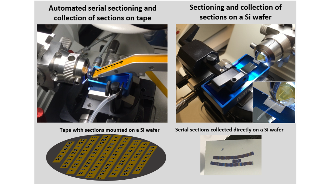
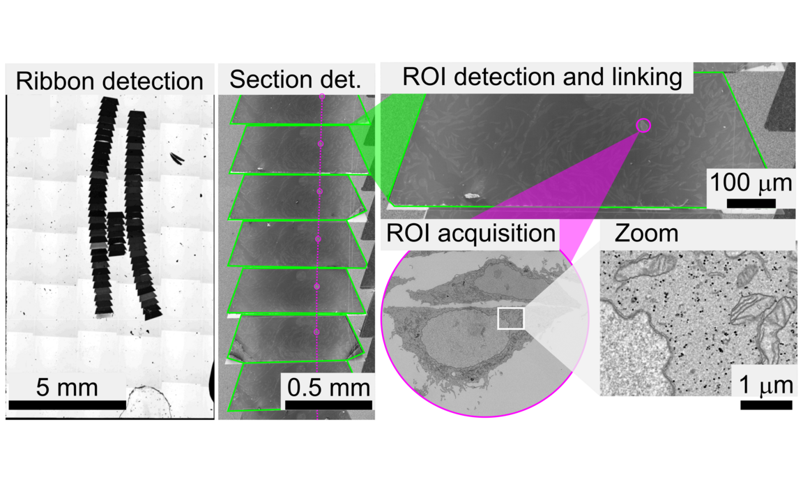
CAT workflow
Unique advantages of CAT
- CAT enables robust registration between the different microscopy modalities, because the same array of ultrathin sections is imaged in the different microscopes.
- CAT, as a non-destructive system compare to en-bloc techniques (FIB-SEM and SB-SEM), permits a certain level of flexibility in experimental design. Additional rounds of immunostaining can be added. Subsequently, upon completion of the electron microscopic imaging, the samples can be revisited again at a later time and reimaged at the SEM at a different magnification, or in a different region. The image stacks on interest can be examined with new questions in mind.
- When targeting rare or specific events within large populations or tissues, CAT is increasingly being recognized as “the method of choice” due to its ability to cover large sample areas (mm-cm).
Video gallery
Interested in CAT services?
If you are interested in CAT services please contact us via this email.